ARG58551
anti-Cathepsin E antibody
anti-Cathepsin E antibody for Western blot and Human
Overview
| Product Description | Rabbit Polyclonal antibody recognizes Cathepsin E |
|---|---|
| Tested Reactivity | Hu |
| Predict Reactivity | Hrs |
| Tested Application | WB |
| Host | Rabbit |
| Clonality | Polyclonal |
| Isotype | IgG |
| Target Name | Cathepsin E |
| Antigen Species | Human |
| Immunogen | Synthetic peptide of Human Cathepsin E. (within the following sequence: SLKKKLRARSQLSEFWKSHNLDMIQFTESCSMDQSAKEPLINYLDMEYFG) |
| Conjugation | Un-conjugated |
| Alternate Names | EC 3.4.23.34; CATE; Cathepsin E |
Application Instructions
| Predict Reactivity Note | Predicted homology based on immunogen sequence: Horse: 79% | ||||
|---|---|---|---|---|---|
| Application Suggestion |
|
||||
| Application Note | * The dilutions indicate recommended starting dilutions and the optimal dilutions or concentrations should be determined by the scientist. | ||||
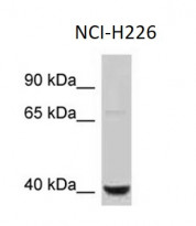
| Positive Control | NCI-H226 whole cell |
Properties
| Form | Liquid |
|---|---|
| Purification | Affinity purified. |
| Buffer | PBS, 0.09% (w/v) Sodium azide and 2% Sucrose. |
| Preservative | 0.09% (w/v) Sodium azide |
| Stabilizer | 2% Sucrose |
| Concentration | Batch dependent: 0.5 - 1 mg/ml |
| Storage Instruction | For continuous use, store undiluted antibody at 2-8°C for up to a week. For long-term storage, aliquot and store at -20°C or below. Storage in frost free freezers is not recommended. Avoid repeated freeze/thaw cycles. Suggest spin the vial prior to opening. The antibody solution should be gently mixed before use. |
| Note | For laboratory research only, not for drug, diagnostic or other use. |
Bioinformation
| Database Links | |
|---|---|
| Gene Symbol | CTSE |
| Gene Full Name | cathepsin E |
| Background | The protein encoded by this gene is a gastric aspartyl protease that functions as a disulfide-linked homodimer. This protease, which is a member of the peptidase A1 family, has a specificity similar to that of pepsin A and cathepsin D. It is an intracellular proteinase that does not appear to be involved in the digestion of dietary protein and is found in highest concentration in the surface of epithelial mucus-producing cells of the stomach. It is the first aspartic proteinase expressed in the fetal stomach and is found in more than half of gastric cancers. It appears, therefore, to be an oncofetal antigen. Transcript variants utilizing alternative polyadenylation signals and two transcript variants encoding different isoforms exist for this gene. [provided by RefSeq, Aug 2015] |
| Function | May have a role in immune function. Probably involved in the processing of antigenic peptides during MHC class II-mediated antigen presentation. May play a role in activation-induced lymphocyte depletion in the thymus, and in neuronal degeneration and glial cell activation in the brain. [UniProt] |
| Calculated MW | 43 kDa |
| PTM | Glycosylated. The nature of the carbohydrate chain varies between cell types. In fibroblasts, the proenzyme contains a high mannose-type oligosaccharide, while the mature enzyme contains a complex-type oligosaccharide. In erythrocyte membranes, both the proenzyme and mature enzyme contain a complex-type oligosaccharide. Two forms are produced by autocatalytic cleavage, form I begins at Ile-54, form II begins at Thr-57. [UniProt] |
Images (1) Click the Picture to Zoom In


