ARG41157
anti-Cathepsin D antibody
anti-Cathepsin D antibody for ICC/IF,IHC-Frozen sections,IHC-Formalin-fixed paraffin-embedded sections,Western blot and Human,Mouse,Rat,Dog,Monkey
Overview
| Product Description | Goat Polyclonal antibody recognizes Cathepsin D |
|---|---|
| Tested Reactivity | Hu, Ms, Rat, Dog, Mk |
| Tested Application | ICC/IF, IHC-Fr, IHC-P, WB |
| Host | Goat |
| Clonality | Polyclonal |
| Isotype | IgG |
| Target Name | Cathepsin D |
| Antigen Species | Human |
| Immunogen | Purified recombinant peptide within aa. 275 to the C-terminus of Human Cathepsin D. |
| Conjugation | Un-conjugated |
| Alternate Names | CPSD; EC 3.4.23.5; HEL-S-130P; CLN10; Cathepsin D |
Application Instructions
| Application Suggestion |
|
||||||||||
|---|---|---|---|---|---|---|---|---|---|---|---|
| Application Note | * The dilutions indicate recommended starting dilutions and the optimal dilutions or concentrations should be determined by the scientist. | ||||||||||
| Positive Control | Human (Jurkat, HT1080, HUH, MDA-MB-231, ARPE19, SH-SY5Y), Dog (MDCK) and Monkey (COS-7) whole cell lysates. |
Properties
| Form | Liquid |
|---|---|
| Purification | Affinity purification with immunogen. |
| Buffer | PBS, 0.05% Sodium azide and 20% Glycerol. |
| Preservative | 0.05% Sodium azide |
| Stabilizer | 20% Glycerol |
| Concentration | 3 mg/ml |
| Storage Instruction | For continuous use, store undiluted antibody at 2-8°C for up to a week. For long-term storage, aliquot and store at -20°C. Storage in frost free freezers is not recommended. Avoid repeated freeze/thaw cycles. Suggest spin the vial prior to opening. The antibody solution should be gently mixed before use. |
| Note | For laboratory research only, not for drug, diagnostic or other use. |
Bioinformation
| Database Links | |
|---|---|
| Gene Symbol | CTSD |
| Gene Full Name | cathepsin D |
| Background | This gene encodes a lysosomal aspartyl protease composed of a dimer of disulfide-linked heavy and light chains, both produced from a single protein precursor. This proteinase, which is a member of the peptidase C1 family, has a specificity similar to but narrower than that of pepsin A. Transcription of this gene is initiated from several sites, including one which is a start site for an estrogen-regulated transcript. Mutations in this gene are involved in the pathogenesis of several diseases, including breast cancer and possibly Alzheimer disease. [provided by RefSeq, Jul 2008] |
| Function | Acid protease active in intracellular protein breakdown. Involved in the pathogenesis of several diseases such as breast cancer and possibly Alzheimer disease. [UniProt] |
| Cellular Localization | Lysosome. Melanosome. Secreted, extracellular space. Note=Identified by mass spectrometry in melanosome fractions from stage I to stage IV. In aortic samples, detected as an extracellular protein loosely bound to the matrix (PubMed:20551380). [UniProt] |
| Calculated MW | 45 kDa |
| PTM | N- and O-glycosylated. Undergoes proteolytic cleavage and activation by ADAM30. As well as the major heavy chain which starts at Leu-169, 2 minor forms starting at Gly-170 and Gly-171 have been identified (PubMed:1426530). An additional form starting at Ala-168 has also been identified (PubMed:27333034). [UniProt] |
Images (5) Click the Picture to Zoom In
-
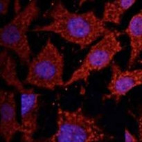
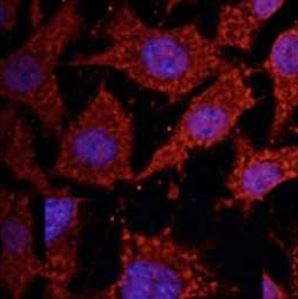
ARG41157 anti-Cathepsin D antibody ICC/IF image
Immunofluorescence: NIH/3T3 cells were fixed with methanol and permeabilized with 0.1% saponin. Cells were stained with ARG41157 anti-Cathepsin D antibody at 1:100 dilution.
-

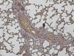
ARG41157 anti-Cathepsin D antibody IHC-P image
Immunohistochemistry: Paraformaldehyde-fixed and paraffin-embedded Mouse lung tissue stained with ARG41157 anti-Cathepsin D antibody at 1:200 dilution.
-
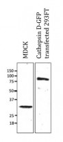
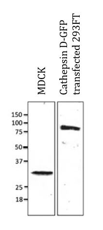
ARG41157 anti-Cathepsin D antibody WB image
Western blot: 100 ug of MDCK cell lysate and 30 ug of transfected 293FT cell lysate stained with ARG41157 anti-Cathepsin D antibody at 1:500 dilution.
-

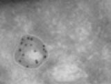
ARG41157 anti-Cathepsin D antibody EM image
Electron microscopy: Immunogold labeling using melanocytes and ARG41157 anti-Cathepsin D antibody.
-
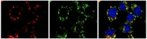
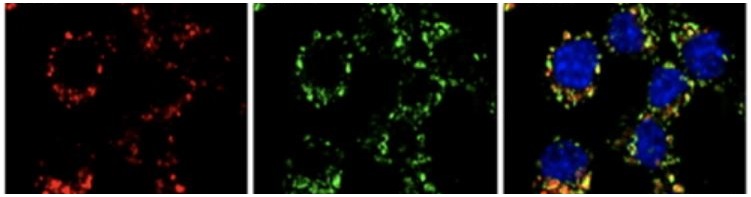
ARG41157 anti-Cathepsin D antibody ICC/IF image
Immunofluorescence: Raw264.7 cells were fixed with PFA and permeabilized with 0.05% saponin. Cells were stained with ARG41157 anti-Cathepsin D antibody (red) at 1:100 dilution. LAMP2 staining (green). nuclear staining (blue).