ARG63969
anti-Catalase antibody
anti-Catalase antibody for IHC-Formalin-fixed paraffin-embedded sections,Western blot and Human,Mouse
Cancer antibody; Cell Biology and Cellular Response antibody; Controls and Markers antibody; Metabolism antibody; Signaling Transduction antibody; Peroxisome Marker antibody
Overview
| Product Description | Goat Polyclonal antibody recognizes Catalase |
|---|---|
| Tested Reactivity | Hu, Ms |
| Predict Reactivity | Dog |
| Tested Application | IHC-P, WB |
| Host | Goat |
| Clonality | Polyclonal |
| Isotype | IgG |
| Target Name | Catalase |
| Antigen Species | Human |
| Immunogen | C-DKYNAEKPKNAIHT |
| Conjugation | Un-conjugated |
| Alternate Names | Catalase; EC 1.11.1.6 |
Application Instructions
| Application Suggestion |
|
||||||
|---|---|---|---|---|---|---|---|
| Application Note | WB: Recommend incubate at RT for 1h. IHC-P: Antigen Retrieval: Steam tissue section in Citrate buffer (pH 6.0). * The dilutions indicate recommended starting dilutions and the optimal dilutions or concentrations should be determined by the scientist. |
Properties
| Form | Liquid |
|---|---|
| Purification | Purified from goat serum by antigen affinity chromatography. |
| Buffer | Tris saline (pH 7.3), 0.02% Sodium azide and 0.5% BSA. |
| Preservative | 0.02% Sodium azide |
| Stabilizer | 0.5% BSA |
| Concentration | 0.5 mg/ml |
| Storage Instruction | For continuous use, store undiluted antibody at 2-8°C for up to a week. For long-term storage, aliquot and store at -20°C or below. Storage in frost free freezers is not recommended. Avoid repeated freeze/thaw cycles. Suggest spin the vial prior to opening. The antibody solution should be gently mixed before use. |
| Note | For laboratory research only, not for drug, diagnostic or other use. |
Bioinformation
| Database Links | |
|---|---|
| Background | This gene encodes catalase, a key antioxidant enzyme in the bodies defense against oxidative stress. Catalase is a heme enzyme that is present in the peroxisome of nearly all aerobic cells. Catalase converts the reactive oxygen species hydrogen peroxide to water and oxygen and thereby mitigates the toxic effects of hydrogen peroxide. Oxidative stress is hypothesized to play a role in the development of many chronic or late-onset diseases such as diabetes, asthma, Alzheimer's disease, systemic lupus erythematosus, rheumatoid arthritis, and cancers. Polymorphisms in this gene have been associated with decreases in catalase activity but, to date, acatalasemia is the only disease known to be caused by this gene. [provided by RefSeq, Oct 2009] |
| Highlight | Related Antibody Duos and Panels: ARG30310 Endosome, Lysosome, Peroxisome Marker Antibody Panel (Catalase, Caveolin1, Clathrin heavy chain, LAMP1) Related products: Catalase antibodies; Catalase ELISA Kits; Catalase Duos / Panels; Anti-Goat IgG secondary antibodies; |
| Research Area | Cancer antibody; Cell Biology and Cellular Response antibody; Controls and Markers antibody; Metabolism antibody; Signaling Transduction antibody; Peroxisome Marker antibody |
| Calculated MW | 60 kDa |
| PTM | The N-terminus is blocked. |
Images (3) Click the Picture to Zoom In
-
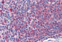
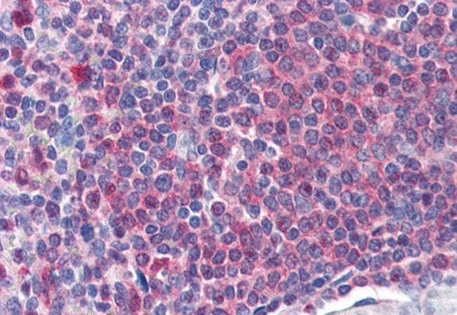
ARG63969 anti-Catalase antibody IHC-P image
Immunohistochemistry: Paraffin-embedded Human spleen tissue. Antigen Retrieval: Steam tissue section in Citrate buffer (pH 6.0). The tissue section was stained with ARG63969 anti-Catalase antibody at 2 µg/ml dilution followed by AP-staining.
-
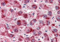
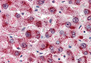
ARG63969 anti-Catalase antibody IHC-P image
Immunohistochemistry: Paraffin embedded Human Liver. (Steamed antigen retrieval with citrate buffer pH 6) stained with ARG63969 anti-Catalase antibody at 2 µg/ml dilution followed by AP-staining.
-
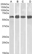
ARG63969 anti-Catalase antibody WB image
Western blot: 35 µg of Human kidney (A), Human liver (B), Mouse kidney (C) and Mouse liver (D) lysates (in RIPA buffer) stained with ARG63969 anti-Catalase antibody at 0.1 µg/ml dilution and incubated at RT for 1 hour.