ARG65765
anti-Caspase 3 (pro) antibody
anti-Caspase 3 (pro) antibody for ICC/IF,IHC-Formalin-fixed paraffin-embedded sections,Western blot and Human,Mouse
Cancer antibody; Cell Biology and Cellular Response antibody; Cell Death antibody; Neuroscience antibody; Apoptosis Marker antibody; Mitochondria/Caspase Dependant Apoptosis Marker antibody

Overview
| Product Description | Rabbit Polyclonal antibody recognizes Caspase 3 (pro) |
|---|---|
| Tested Reactivity | Hu, Ms |
| Tested Application | ICC/IF, IHC-P, WB |
| Specificity | This antibody recognizes full length Caspase 3 only. |
| Host | Rabbit |
| Clonality | Polyclonal |
| Isotype | IgG |
| Target Name | Caspase 3 (pro) |
| Antigen Species | Human |
| Immunogen | Synthetic peptide derived from Human pro Caspase 3. |
| Conjugation | Un-conjugated |
| Alternate Names | CASP3; Caspase 3; Apopain; CPP32; CPP32B; Caspase 3, Apoptosis-Related Cysteine Peptidase; Caspase 3, Apoptosis-Related Cysteine Protease; SREBP Cleavage Activity 1; Cysteine Protease CPP32; Protein Yama; EC 3.4.22.56; Caspase-3; CASP-3; CPP-32; SCA-1; Yama; PARP Cleavage Protease; Procaspase3; EC 3.4.22 |
Application Instructions
| Application Suggestion |
|
||||||||
|---|---|---|---|---|---|---|---|---|---|
| Application Note | * The dilutions indicate recommended starting dilutions and the optimal dilutions or concentrations should be determined by the scientist. | ||||||||
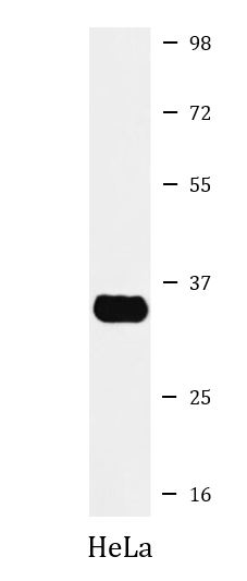
| Positive Control | HeLa | ||||||||
| Observed Size | ~ 34 kDa |
Properties
| Form | Liquid |
|---|---|
| Purification | Affinity purified. |
| Buffer | PBS (pH 7.4), 150 mM NaCl, 0.02% Sodium azide and 50% Glycerol. |
| Preservative | 0.02% Sodium azide |
| Stabilizer | 50% Glycerol |
| Storage Instruction | For continuous use, store undiluted antibody at 2-8°C for up to a week. For long-term storage, aliquot and store at -20°C. Storage in frost free freezers is not recommended. Avoid repeated freeze/thaw cycles. Suggest spin the vial prior to opening. The antibody solution should be gently mixed before use. |
| Note | For laboratory research only, not for drug, diagnostic or other use. |
Bioinformation
| Database Links | |
|---|---|
| Gene Symbol | CASP3 |
| Gene Full Name | caspase 3, apoptosis-related cysteine peptidase |
| Background | The protein encoded by this gene is a cysteine-aspartic acid protease that plays a central role in the execution-phase of cell apoptosis. The encoded protein cleaves and inactivates poly(ADP-ribose) polymerase while it cleaves and activates sterol regulatory element binding proteins as well as caspases 6, 7, and 9. This protein itself is processed by caspases 8, 9, and 10. It is the predominant caspase involved in the cleavage of amyloid-beta 4A precursor protein, which is associated with neuronal death in Alzheimer's disease. [provided by RefSeq, Aug 2017] |
| Function | Cleaves XRCC4 and phospholipid scramblase proteins XKR4, XKR8 and XKR9, leading to promote phosphatidylserine exposure on apoptotic cell surface. [UniProt] |
| Cellular Localization | Cytoplasm. [UniProt] |
| Highlight | Related Antibody Duos and Panels: ARG30105 Apoptosis Marker Antibody Duo (Caspase3, PARP) ARG30110 Mitochondria/Caspase dependant Apoptosis Antibody Panel (Caspase3, Caspase9, Cytochrome c, PARP) (WB) Related products: Caspase 3 antibodies; Caspase 3 Duos / Panels; |
| Research Area | Cancer antibody; Cell Biology and Cellular Response antibody; Cell Death antibody; Neuroscience antibody; Apoptosis Marker antibody; Mitochondria/Caspase Dependant Apoptosis Marker antibody |
| Calculated MW | 32 kDa |
| PTM | Acetylation, Phosphoprotein, S-nitrosylation, Zymogen. [UniProt] |
Images (2) Click the Picture to Zoom In
-

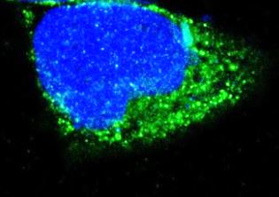
ARG65765 anti-Caspase 3 (pro) antibody ICC/IF image
Immunofluorescence: A673 cells stained with ARG65765 anti-Caspase 3 (pro) antibody.
-
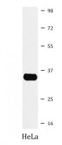
ARG65765 anti-Caspase 3 (pro) antibody WB image
Western blot: HeLa cell lysate stained with ARG65765 anti-Caspase 3 (pro) antibody.
Customer's Feedback
Good
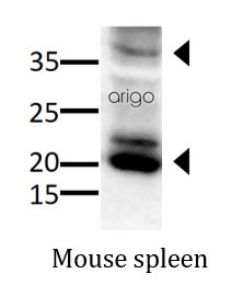
Review for anti-Caspase 3 (pro) antibody
Application:WB
Sample:Mouse spleen
Sample Loading Amount:30 µg
Primary Antibody Dilution Factor:1:500
Primary Antibody Incubation Time:overnight
Primary Antibody Incubation Temperature:4 ºC

Specific References
Cobalt protoporphyrin promotes human keratinocyte migration under hyperglycemic conditions
WB / Human


