ARG43230
anti-Caspase 3 antibody [8B6]
anti-Caspase 3 antibody [8B6] for Flow cytometry,IHC-Formalin-fixed paraffin-embedded sections,Western blot and Human
Cancer antibody; Cell Biology and Cellular Response antibody; Cell Death antibody; Neuroscience antibody; Apoptosis Marker antibody; Mitochondria/Caspase Dependant Apoptosis Marker antibody
Overview
| Product Description | Mouse Monoclonal antibody [8B6] recognizes Caspase 3 |
|---|---|
| Tested Reactivity | Hu |
| Tested Application | FACS, IHC-P, WB |
| Host | Mouse |
| Clonality | Monoclonal |
| Clone | 8B6 |
| Isotype | IgG1 |
| Target Name | Caspase 3 |
| Antigen Species | Human |
| Immunogen | Recombinant protein corresponding to T67-D175 of Human Caspase 3. |
| Conjugation | Un-conjugated |
| Alternate Names | CASP3; Caspase 3; Apopain; CPP32; CPP32B; Caspase 3, Apoptosis-Related Cysteine Peptidase; Caspase 3, Apoptosis-Related Cysteine Protease; SREBP Cleavage Activity 1; Cysteine Protease CPP32; Protein Yama; EC 3.4.22.56; Caspase-3; CASP-3; CPP-32; SCA-1; Yama; PARP Cleavage Protease; Procaspase3; EC 3.4.22 |
Application Instructions
| Application Suggestion |
|
||||||||
|---|---|---|---|---|---|---|---|---|---|
| Application Note | IHC-P: Antigen Retrieval: Heat mediation was performed in EDTA buffer (pH 8.0). * The dilutions indicate recommended starting dilutions and the optimal dilutions or concentrations should be determined by the scientist. |
||||||||
| Observed Size | ~ 35 kDa |
Properties
| Form | Liquid |
|---|---|
| Purification | Affinity purification with immunogen. |
| Buffer | 0.2% Na2HPO4, 0.9% NaCl, 0.05% Sodium azide and 4% Trehalose. |
| Preservative | 0.05% Sodium azide |
| Stabilizer | 4% Trehalose |
| Concentration | 0.5 mg/ml |
| Storage Instruction | For continuous use, store undiluted antibody at 2-8°C for up to a week. For long-term storage, aliquot and store at -20°C or below. Storage in frost free freezers is not recommended. Avoid repeated freeze/thaw cycles. Suggest spin the vial prior to opening. The antibody solution should be gently mixed before use. |
| Note | For laboratory research only, not for drug, diagnostic or other use. |
Bioinformation
| Database Links | |
|---|---|
| Gene Symbol | CASP3 |
| Gene Full Name | caspase 3, apoptosis-related cysteine peptidase |
| Background | The protein encoded by this gene is a cysteine-aspartic acid protease that plays a central role in the execution-phase of cell apoptosis. The encoded protein cleaves and inactivates poly(ADP-ribose) polymerase while it cleaves and activates sterol regulatory element binding proteins as well as caspases 6, 7, and 9. This protein itself is processed by caspases 8, 9, and 10. It is the predominant caspase involved in the cleavage of amyloid-beta 4A precursor protein, which is associated with neuronal death in Alzheimer's disease. [provided by RefSeq, Aug 2017] |
| Function | Cleaves XRCC4 and phospholipid scramblase proteins XKR4, XKR8 and XKR9, leading to promote phosphatidylserine exposure on apoptotic cell surface. [UniProt] |
| Cellular Localization | Cytoplasm. [UniProt] |
| Highlight | Related Antibody Duos and Panels: ARG30105 Apoptosis Marker Antibody Duo (Caspase3, PARP) ARG30110 Mitochondria/Caspase dependant Apoptosis Antibody Panel (Caspase3, Caspase9, Cytochrome c, PARP) (WB) Related products: Caspase 3 antibodies; Caspase 3 Duos / Panels; Anti-Mouse IgG secondary antibodies; |
| Research Area | Cancer antibody; Cell Biology and Cellular Response antibody; Cell Death antibody; Neuroscience antibody; Apoptosis Marker antibody; Mitochondria/Caspase Dependant Apoptosis Marker antibody |
| Calculated MW | 32 kDa |
| PTM | Acetylation, Phosphoprotein, S-nitrosylation, Zymogen. [UniProt] |
Images (5) Click the Picture to Zoom In
-

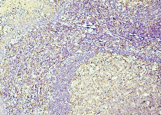
ARG43230 anti-Caspase 3 antibody [8B6] IHC-P image
Immunohistochemistry: Paraffin-embedded Human tonsil tissue. Antigen Retrieval: Heat mediation was performed in EDTA buffer (pH 8.0). The tissue section was blocked with 10% goat serum. The tissue section was then stained with ARG43230 anti-Caspase 3 antibody [8B6] at 1 µg/ml dilution, overnight at 4°C.
-
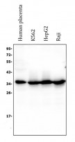
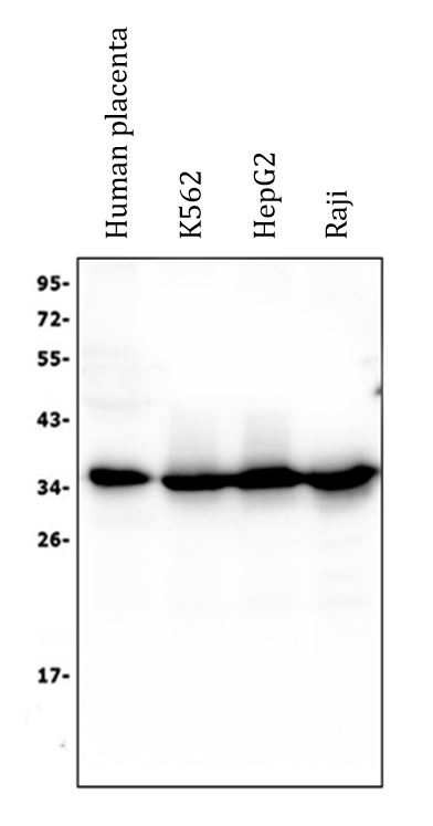
ARG43230 anti-Caspase 3 antibody [8B6] WB image
Western blot: 50 µg of sample under reducing conditions. Human placenta, K562, HepG2 and Raji whole cell lysates stained with ARG43230 anti-Caspase 3 antibody [8B6] at 0.5 µg/ml dilution, overnight at 4°C.
-
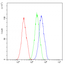
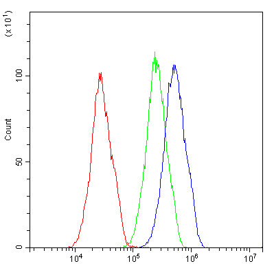
ARG43230 anti-Caspase 3 antibody [8B6] FACS image
Flow Cytometry: HepG2 cells were blocked with 10% normal goat serum and then stained with ARG43230 anti-Caspase 3 antibody [8B6] (blue) at 1 µg/10^6 cells for 30 min at 20°C, followed by incubation with DyLight®488 labelled secondary antibody. Isotype control antibody (green) was mouse IgG (1 µg/10^6 cells) used under the same conditions. Unlabelled sample (red) was also used as a control.
-
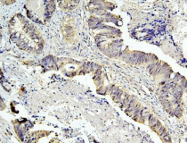
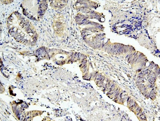
ARG43230 anti-Caspase 3 antibody [8B6] IHC-P image
Immunohistochemistry: Paraffin-embedded Human intestinal cancer tissue. Antigen Retrieval: Heat mediation was performed in EDTA buffer (pH 8.0). The tissue section was blocked with 10% goat serum. The tissue section was then stained with ARG43230 anti-Caspase 3 antibody [8B6] at 1 µg/ml dilution, overnight at 4°C.
-
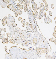
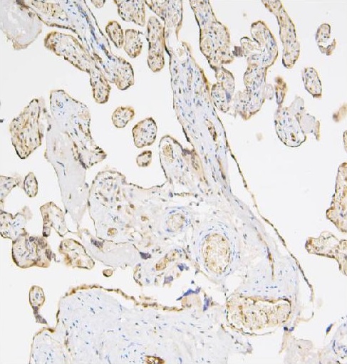
ARG43230 anti-Caspase 3 antibody [8B6] IHC-P image
Immunohistochemistry: Paraffin-embedded Human placenta tissue. Antigen Retrieval: Heat mediation was performed in EDTA buffer (pH 8.0). The tissue section was blocked with 10% goat serum. The tissue section was then stained with ARG43230 anti-Caspase 3 antibody [8B6] at 1 µg/ml dilution, overnight at 4°C.