ARG10704
anti-Calretinin antibody
anti-Calretinin antibody for ICC/IF,IHC-Frozen sections,Western blot and Human,Mouse,Rat,Cow
Overview
| Product Description | Rabbit Polyclonal antibody recognizes Calretinin |
|---|---|
| Tested Reactivity | Hu, Ms, Rat, Cow |
| Tested Application | ICC/IF, IHC-Fr, WB |
| Host | Rabbit |
| Clonality | Polyclonal |
| Isotype | IgG |
| Target Name | Calretinin |
| Antigen Species | Human |
| Immunogen | Full-length recombinant Human protein. |
| Conjugation | Un-conjugated |
| Alternate Names | CAB29; CR; CAL2; 29 kDa calbindin; Calretinin |
Application Instructions
| Application Suggestion |
|
||||||||
|---|---|---|---|---|---|---|---|---|---|
| Application Note | * The dilutions indicate recommended starting dilutions and the optimal dilutions or concentrations should be determined by the scientist. |
Properties
| Form | Liquid |
|---|---|
| Purification | Crude rabbit serum. |
| Buffer | Serum. |
| Storage Instruction | For continuous use, store undiluted antibody at 2-8°C for up to a week. For long-term storage, aliquot and store at -20°C or below. Storage in frost free freezers is not recommended. Avoid repeated freeze/thaw cycles. Suggest spin the vial prior to opening. The antibody solution should be gently mixed before use. |
| Note | For laboratory research only, not for drug, diagnostic or other use. |
Bioinformation
| Database Links | |
|---|---|
| Gene Symbol | CALB2 |
| Gene Full Name | calbindin 2 |
| Background | This gene encodes an intracellular calcium-binding protein belonging to the troponin C superfamily. Members of this protein family have six EF-hand domains which bind calcium. This protein plays a role in diverse cellular functions, including message targeting and intracellular calcium buffering. It also functions as a modulator of neuronal excitability, and is a diagnostic marker for some human diseases, including Hirschsprung disease and some cancers. Alternative splicing results in multiple transcript variants. [provided by RefSeq, Jun 2010] |
| Function | Calretinin is a calcium-binding protein which is abundant in auditory neurons. [UniProt] |
| Calculated MW | 32 kDa |
Images (6) Click the Picture to Zoom In
-
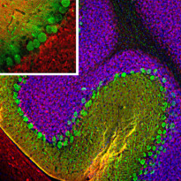
ARG10704 anti-Calretinin antibody IHC-Fr image
Immunohistochemistry: Frozen section of Rat cerebellum stained with ARG10704 anti-Calretinin antibody (red) at 1:5000 dilution and costained with Mouse mAb to calbindin (green) at 1:1000 dilution. (Sample preparation: Following transcardial perfusion of Rat with 4% paraformaldehyde, brain was post fixed for 24 hours, cut to 45 µM, and free-floating sections were stained with the above antibodies.)
The calretinin antibody stains interneurons predominantly in the molecular layer, while the calbindin antibody strongly labels the dendrites and perikarya of Purkinje cells in the molecular layer of the cerebellum.
-
ARG10704 anti-Calretinin antibody IHC-Fr image
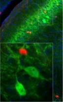
Immunohistochemistry: Frozen sections of adult Mouse brain (45 µM; fixed by transcardial perfusion with 4% paraformaldehyde) across motor cortex was co-stained with ARG10704 anti-Calretinin antibody (red) and a Mouse monoclonal 1B7 to Fox3 / NeuN (green).
-
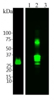
ARG10704 anti-Calretinin antibody WB image
Western blot: Left: 20 µg of Rat brain lysates stained with ARG10704 anti-Calretinin antibody at 1:5000 dilution. Right: 0.2 µg of Human 1) parvalbumin, 2) calretinin, and 3) calbindin recombinant proteins.
-
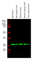
ARG10704 anti-Calretinin antibody WB image
Western blot: Rat brain, Rat spinal cord, Mouse brain, Mouse spinal cord and Cow spinal cord lysates stained with ARG10704 anti-Calretinin antibody (green) at 1:10000 dilution.
-
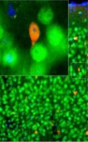
ARG10704 anti-Calretinin antibody IHC-Fr image
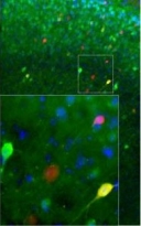
Immunohistochemistry: Frozen sections of adult Mouse brain across visual cortex was stained with ARG10704 anti-Calretinin antibody (red) and co-stained with our chicken polyclonal antibody to calbindin (green). Calretinin and calbindin label different population of neurons in the brain. As a result, most cells were stained with one of the two antibodies and appear to be either red or green. However in visual cortex, a few cells express both proteins and appear to be yellow.
-
ARG10704 anti-Calretinin antibody IHC-Fr image
Immunohistochemistry: Frozen sections of adult Rat brain (45 µM; fixed by transcardial perfusion with 4% paraformaldehyde) across hippocampal CA1 region was stained with ARG10704 anti-Calretinin antibody (red) and our Mouse monoclonal 3C9 to parvalbumin (green). The two antibodies stain distinct subsets of interneurons in the pyramidal layer and the positively labeled cells appear to be either red or green. Blue is a Hoechst staining that labels DNA.


