ARG10680
anti-Calretinin antibody
anti-Calretinin antibody for ICC/IF,IHC-Frozen sections,Western blot and Human,Mouse,Rat,Cow
Overview
| Product Description | Chicken Polyclonal antibody recognizes Calretinin |
|---|---|
| Tested Reactivity | Hu, Ms, Rat, Cow |
| Tested Application | ICC/IF, IHC-Fr, WB |
| Host | Chicken |
| Clonality | Polyclonal |
| Isotype | IgY |
| Target Name | Calretinin |
| Antigen Species | Human |
| Immunogen | Full-length recombinant Human Calretinin protein. |
| Conjugation | Un-conjugated |
| Alternate Names | CAB29; CR; CAL2; 29 kDa calbindin; Calretinin |
Application Instructions
| Application Suggestion |
|
||||||||
|---|---|---|---|---|---|---|---|---|---|
| Application Note | * The dilutions indicate recommended starting dilutions and the optimal dilutions or concentrations should be determined by the scientist. |
Properties
| Form | Liquid |
|---|---|
| Buffer | PBS and 0.02% Sodium azide. |
| Preservative | 0.02% Sodium azide |
| Storage Instruction | For continuous use, store undiluted antibody at 2-8°C for up to a week. For long-term storage, aliquot and store at -20°C or below. Storage in frost free freezers is not recommended. Avoid repeated freeze/thaw cycles. Suggest spin the vial prior to opening. The antibody solution should be gently mixed before use. |
| Note | For laboratory research only, not for drug, diagnostic or other use. |
Bioinformation
| Database Links | |
|---|---|
| Gene Symbol | CALB2 |
| Gene Full Name | calbindin 2 |
| Background | This gene encodes an intracellular calcium-binding protein belonging to the troponin C superfamily. Members of this protein family have six EF-hand domains which bind calcium. This protein plays a role in diverse cellular functions, including message targeting and intracellular calcium buffering. It also functions as a modulator of neuronal excitability, and is a diagnostic marker for some human diseases, including Hirschsprung disease and some cancers. Alternative splicing results in multiple transcript variants. [provided by RefSeq, Jun 2010] |
| Function | Calretinin is a calcium-binding protein which is abundant in auditory neurons. [UniProt] |
| Calculated MW | 32 kDa |
Images (5) Click the Picture to Zoom In
-
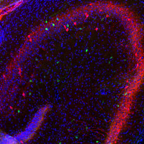
ARG10680 anti-Calretinin antibody IHC-Fr image
Immunohistochemistry: Frozen section of Rat hippocampus stained with ARG10680 anti-Calretinin antibody (green) at 1:1000 dilution and costained with ARG10725 anti-Parvalbumin antibody [3C9] (red) at 1:1000 dilution. (Sample preparation: Following transcardial perfusion of Rat with 4% paraformaldehyde, brain was post fixed for 24 hours, cut to 45 µM, and free-floating sections were stained with the above antibodies.)
-
ARG10680 anti-Calretinin antibody IHC-Fr image
Immunohistochemistry: Frozen sections of adult Rat brain hippocampus (45 µM; fixed by transcardial perfusion with 4% paraformaldehyde) was stained with ARG10680 anti-Calretinin antibody at 1:1000 (red), and co-stained with our rabbit anti-MeCP2 antibody (green). Calretinin labels a subset of hippocampal interneurons, which also express MeCP2 in the nucleus to give a yellow color.
-
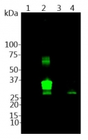
ARG10680 anti-Calretinin antibody WB image
Western blot: 1) parvalbumin, 2) calretinin, 3) calbindin recombinant proteins, and 4) Rat brain lysates was stained with ARG10680 anti-Calretinin antibody at 1:1000 dilution.
-
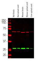
ARG10680 anti-Calretinin antibody WB image
Western blot: Rat brain, Rat spinal cord, Mouse brain, Mouse spinal cord and Cow spinal cord lysates stained with ARG10680 anti-Calretinin antibody (green) at 1:1000 dilution. The same blot was simultaneously stained with Mouse mAb to alpha-nternexin (red) at 1:10000 dilution.
-
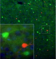
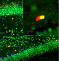
ARG10680 anti-Calretinin antibody IHC-Fr image
Immunohistochemistry: Frozen sections of adult Rat cortex was stained with ARG10680 anti-Calretinin antibody (red) and co-stained with Mouse anti-calbindin antibody (green). Each antibody specifically labels a subset of interneurons (i.e., calretinin-positive or calbindin-postive) that express each marker exclusively. Inset is a high-magnification image of the boxed area. Blue is DAPI staining that labels DNA.


