ARG40287
anti-Calbindin antibody
anti-Calbindin antibody for IHC-Frozen sections,IHC-Formalin-fixed paraffin-embedded sections,Western blot and Human,Mouse,Rat
Overview
| Product Description | Goat Polyclonal antibody recognizes Calbindin |
|---|---|
| Tested Reactivity | Hu, Ms, Rat |
| Predict Reactivity | Cow, Dog, Pig |
| Tested Application | IHC-Fr, IHC-P, WB |
| Host | Goat |
| Clonality | Polyclonal |
| Isotype | IgG |
| Target Name | Calbindin |
| Antigen Species | Human |
| Immunogen | Synthetic peptide around the internal region of Human Calbindin. (NP_004920.1. C-KTFVDQYGQRDDGK) |
| Conjugation | Un-conjugated |
| Alternate Names | Vitamin D-dependent calcium-binding protein, avian-type; Calbindin; CALB; Calbindin D28; D-28K |
Application Instructions
| Application Suggestion |
|
||||||||
|---|---|---|---|---|---|---|---|---|---|
| Application Note | WB: Recommend incubate at RT for 1h. IHC-P: Antigen Retrieval: Steam tissue section in Citrate buffer (pH 6.0) at 80°C for 30 min. * The dilutions indicate recommended starting dilutions and the optimal dilutions or concentrations should be determined by the scientist. |
||||||||
| Observed Size | ~ 26 kDa |
Properties
| Form | Liquid |
|---|---|
| Purification | Affinity purified |
| Buffer | Tris saline (pH 7.3), 0.02% Sodium azide and 0.5% BSA. |
| Preservative | 0.02% Sodium azide |
| Stabilizer | 0.5% BSA |
| Concentration | 0.5 mg/ml |
| Storage Instruction | For continuous use, store undiluted antibody at 2-8°C for up to a week. For long-term storage, aliquot and store at -20°C or below. Storage in frost free freezers is not recommended. Avoid repeated freeze/thaw cycles. Suggest spin the vial prior to opening. The antibody solution should be gently mixed before use. |
| Note | For laboratory research only, not for drug, diagnostic or other use. |
Bioinformation
| Database Links | |
|---|---|
| Gene Symbol | CALB1 |
| Gene Full Name | calbindin 1, 28kDa |
| Background | The protein encoded by this gene is a member of the calcium-binding protein superfamily that includes calmodulin and troponin C. Originally described as a 27 kDa protein, it is now known to be a 28 kDa protein. It contains four active calcium-binding domains, and has two modified domains that are thought to have lost their calcium binding capability. This protein is thought to buffer entry of calcium upon stimulation of glutamate receptors. Depletion of this protein was noted in patients with Huntington disease. [provided by RefSeq, Jan 2015] |
| Function | Buffers cytosolic calcium. May stimulate a membrane Ca(2+)-ATPase and a 3',5'-cyclic nucleotide phosphodiesterase. [UniProt] |
| Calculated MW | 30 kDa |
Images (4) Click the Picture to Zoom In
-

ARG40287 anti-Calbindin antibody IHC-Fr image
Immunohistochemistry: PFA-perfused cryosection of Human hypothalamus tissue stained with ARG40287 anti-Calbindin antibody at 0.1 µg/ml dilution.
-
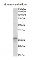
ARG40287 anti-Calbindin antibody WB image
Western blot: 35 µg of Human cerebellum lysate (in RIPA buffer) stained with ARG40287 anti-Calbindin antibody at 0.03 µg/ml dilution and incubated at RT for 1 hour.
-
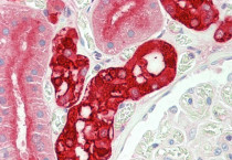
ARG40287 anti-Calbindin antibody IHC-P image
Immunohistochemistry: Paraffin-embedded Human kidney stained with ARG40287 anti-Calbindin antibody at 5 µg/ml dilution. Antigen Retrieval: Steam tissue section in Citrate buffer (pH 6.0).
-
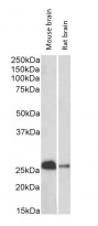
ARG40287 anti-Calbindin antibody WB image
Western blot: 35 µg of Mouse brain and Rat brain lysates (in RIPA buffer) stained with ARG40287 anti-Calbindin antibody at 0.1 µg/ml dilution and incubated at RT for 1 hour.


