ARG54726
anti-CYP3A4 antibody
anti-CYP3A4 antibody for Flow cytometry,IHC-Formalin-fixed paraffin-embedded sections,Western blot and Human
Cell Biology and Cellular Response antibody; Metabolism antibody; Signaling Transduction antibody
Overview
| Product Description | Rabbit Polyclonal antibody recognizes CYP3A4 |
|---|---|
| Tested Reactivity | Hu |
| Tested Application | FACS, IHC-P, WB |
| Host | Rabbit |
| Clonality | Polyclonal |
| Isotype | IgG |
| Target Name | CYP3A4 |
| Antigen Species | Human |
| Immunogen | KLH-conjugated synthetic peptide corresponding to aa. 228-255 (Center) of Human CYP3A4 (NP_001189784.1). |
| Conjugation | Un-conjugated |
| Alternate Names | EC 1.14.13.-; Nifedipine oxidase; P450PCN1; EC 1.14.13.32; Taurochenodeoxycholate 6-alpha-hydroxylase; Albendazole sulfoxidase; EC 1.14.13.97; Cytochrome P450 NF-25; CYPIIIA4; Cytochrome P450 HLp; CYPIIIA3; EC 1.14.13.157; Cytochrome P450 3A4; Cytochrome P450 3A3; P450C3; EC 1.14.13.67; Quinine 3-monooxygenase; Albendazole monooxygenase; CP34; CYP3A3; CP33; 1,8-cineole 2-exo-monooxygenase; NF-25; HLP; Cytochrome P450-PCN1; CYP3A |
Application Instructions
| Application Suggestion |
|
||||||||
|---|---|---|---|---|---|---|---|---|---|
| Application Note | * The dilutions indicate recommended starting dilutions and the optimal dilutions or concentrations should be determined by the scientist. | ||||||||
| Positive Control | HepG2 |
Properties
| Purification | This antibody is prepared by Saturated Ammonium Sulfate (SAS) precipitation followed by dialysis against PBS. |
|---|---|
| Buffer | PBS and 0.09% (W/V) Sodium azide |
| Preservative | 0.09% (W/V) Sodium azide |
| Storage Instruction | For continuous use, store undiluted antibody at 2-8°C for up to a week. For long-term storage, aliquot and store at -20°C or below. Storage in frost free freezers is not recommended. Avoid repeated freeze/thaw cycles. Suggest spin the vial prior to opening. The antibody solution should be gently mixed before use. |
| Note | For laboratory research only, not for drug, diagnostic or other use. |
Bioinformation
| Database Links | |
|---|---|
| Gene Symbol | CYP3A4 |
| Gene Full Name | cytochrome P450, family 3, subfamily A, polypeptide 4 |
| Background | This gene encodes a member of the cytochrome P450 superfamily of enzymes. The cytochrome P450 proteins are monooxygenases that catalyze many reactions involved in drug metabolism and synthesis of cholesterol, steroids and other lipids. This protein localizes to the endoplasmic reticulum and its expression is induced by glucocorticoids and some pharmacological agents. This enzyme is involved in the metabolism of approximately half the drugs in use today, including acetaminophen, codeine, cyclosporin A, diazepam and erythromycin. The enzyme also metabolizes some steroids and carcinogens. This gene is part of a cluster of cytochrome P450 genes on chromosome 7q21.1. Previously another CYP3A gene, CYP3A3, was thought to exist; however, it is now thought that this sequence represents a transcript variant of CYP3A4. Alternatively spliced transcript variants encoding different isoforms have been identified. [provided by RefSeq, Feb 2011] |
| Function | Cytochromes P450 are a group of heme-thiolate monooxygenases. In liver microsomes, this enzyme is involved in an NADPH-dependent electron transport pathway. It performs a variety of oxidation reactions (e.g. caffeine 8-oxidation, omeprazole sulphoxidation, midazolam 1'-hydroxylation and midazolam 4- hydroxylation) of structurally unrelated compounds, including steroids, fatty acids, and xenobiotics. Acts as a 1,8-cineole 2- exo-monooxygenase. The enzyme also hydroxylates etoposide. [From Uniprot] |
| Cellular Localization | Endoplasmic reticulum membrane; Single-pass membrane protein. Microsome membrane; Single-pass membrane protein |
| Research Area | Cell Biology and Cellular Response antibody; Metabolism antibody; Signaling Transduction antibody |
| Calculated MW | 57 kDa |
| PTM | Polyubiquitinated in the presence of AMFR and UBE2G1 and also STUB1/CHIP and UBE2D1 (in vitro). |
Images (3) Click the Picture to Zoom In
-

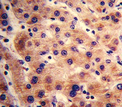
ARG54726 anti-CYP3A4 antibody IHC-P image
Immunohistochemistry: Formalin-fixed and paraffin-embedded Human liver tissue stained with ARG54726 anti-CYP3A4 antibody.
-

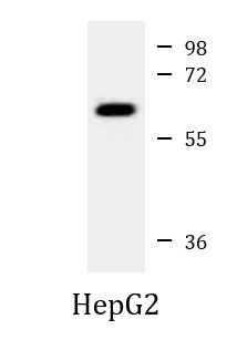
ARG54726 anti-CYP3A4 antibody WB image
Western blot: 35 µg of HepG2 cell lysate stained with ARG54726 anti-CYP3A4 antibody at 1:1000 dilution.
-

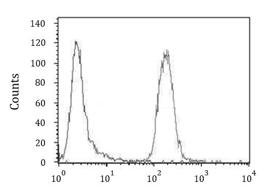
ARG54726 anti-CYP3A4 antibody FACS image
Flow Cytometry: CEM cells stained with ARG54726 anti-CYP3A4 antibody (right histogram) or without primary antibody control (left histogram).