ARG42303
anti-CXCR4 antibody [12G5] (APC)
anti-CXCR4 antibody [12G5] (APC) for Flow cytometry and Human,Primates
Overview
| Product Description | APC-conjugated Mouse Monoclonal antibody [12G5] recognizes CXCR4 |
|---|---|
| Tested Reactivity | Hu, NHuPrm |
| Tested Application | FACS |
| Specificity | The mouse monoclonal antibody 12G5 recognizes an extracellular epitope of CD184, a 45 kDa G-protein-linked CXC chemokine receptor widely expressed on blood and tissue cells. |
| Host | Mouse |
| Clonality | Monoclonal |
| Clone | 12G5 |
| Isotype | IgG2a, kappa |
| Target Name | CXCR4 |
| Antigen Species | Human |
| Immunogen | CP-MAC-infected Sup-T1 cells. |
| Conjugation | APC |
| Alternate Names | Lipopolysaccharide-associated protein 3; LAP-3; LAP3; Stromal cell-derived factor 1 receptor; Leukocyte-derived seven transmembrane domain receptor; WHIMS; NPY3R; SDF-1 receptor; Fusin; LPS-associated protein 3; HM89; HSY3RR; FB22; NPYR; CD antigen CD184; LCR1; NPYY3R; WHIM; D2S201E; C-X-C chemokine receptor type 4; LESTR; CXC-R4; CD184; NPYRL; CXCR-4 |
Application Instructions
| Application Suggestion |
|
||||
|---|---|---|---|---|---|
| Application Note | * The dilutions indicate recommended starting dilutions and the optimal dilutions or concentrations should be determined by the scientist. |
Properties
| Form | Liquid |
|---|---|
| Purification | Purified |
| Buffer | PBS and 15 mM Sodium azide. |
| Preservative | 15 mM Sodium azide |
| Storage Instruction | Aliquot and store in the dark at 2-8°C. Keep protected from prolonged exposure to light. Avoid repeated freeze/thaw cycles. Suggest spin the vial prior to opening. The antibody solution should be gently mixed before use. |
| Note | For laboratory research only, not for drug, diagnostic or other use. |
Bioinformation
| Database Links | |
|---|---|
| Gene Symbol | CXCR4 |
| Gene Full Name | chemokine (C-X-C motif) receptor 4 |
| Background | This gene encodes a CXC chemokine receptor specific for stromal cell-derived factor-1. The protein has 7 transmembrane regions and is located on the cell surface. It acts with the CD4 protein to support HIV entry into cells and is also highly expressed in breast cancer cells. Mutations in this gene have been associated with WHIM (warts, hypogammaglobulinemia, infections, and myelokathexis) syndrome. Alternate transcriptional splice variants, encoding different isoforms, have been characterized. [provided by RefSeq, Jul 2008] |
| Function | Receptor for the C-X-C chemokine CXCL12/SDF-1 that transduces a signal by increasing intracellular calcium ion levels and enhancing MAPK1/MAPK3 activation (PubMed:10452968, PubMed:28978524, PubMed:18799424, PubMed:24912431). Involved in the AKT signaling cascade (PubMed:24912431). Plays a role in regulation of cell migration, e.g. during wound healing (PubMed:28978524). Acts as a receptor for extracellular ubiquitin; leading to enhanced intracellular calcium ions and reduced cellular cAMP levels (PubMed:20228059). Binds bacterial lipopolysaccharide (LPS) et mediates LPS-induced inflammatory response, including TNF secretion by monocytes (PubMed:11276205). Involved in hematopoiesis and in cardiac ventricular septum formation. Also plays an essential role in vascularization of the gastrointestinal tract, probably by regulating vascular branching and/or remodeling processes in endothelial cells. Involved in cerebellar development. In the CNS, could mediate hippocampal-neuron survival (By similarity). (Microbial infection) Acts as a coreceptor (CD4 being the primary receptor) for human immunodeficiency virus-1/HIV-1 X4 isolates and as a primary receptor for some HIV-2 isolates. Promotes Env-mediated fusion of the virus (PubMed:8849450, PubMed:8929542, PubMed:9427609, PubMed:10074122, PubMed:10756055). [UniProt] |
| Cellular Localization | Cell membrane; Multi-pass membrane protein. Cell junction. Early endosome. Late endosome. Lysosome. Note=In unstimulated cells, diffuse pattern on plasma membrane. On agonist stimulation, colocalizes with ITCH at the plasma membrane where it becomes ubiquitinated. In the presence of antigen, distributes to the immunological synapse forming at the T-cell-APC contact area, where it localizes at the peripheral and distal supramolecular activation cluster (SMAC). [UniProt] |
| Calculated MW | 40 kDa |
| PTM | Phosphorylated on agonist stimulation. Rapidly phosphorylated on serine and threonine residues in the C-terminal. Phosphorylation at Ser-324 and Ser-325 leads to recruitment of ITCH, ubiquitination and protein degradation. Ubiquitinated by ITCH at the cell membrane on agonist stimulation. The ubiquitin-dependent mechanism, endosomal sorting complex required for transport (ESCRT), then targets CXCR4 for lysosomal degradation. This process is dependent also on prior Ser-/Thr-phosphorylation in the C-terminal of CXCR4. Also binding of ARRB1 to STAM negatively regulates CXCR4 sorting to lysosomes though modulating ubiquitination of SFR5S. Sulfation on Tyr-21 is required for efficient binding of CXCL12/SDF-1alpha and promotes its dimerization. Tyr-7 and Tyr-12 are sulfated in a sequential manner after Tyr-21 is almost fully sulfated, with the binding affinity for CXCL12/SDF-1alpha increasing with the number of sulfotyrosines present. Sulfotyrosines Tyr-7 and Tyr-12 occupy clefts on opposing CXCL12 subunits, thus bridging the CXCL12 dimer interface and promoting CXCL12 dimerization. O- and N-glycosylated. Asn-11 is the principal site of N-glycosylation. There appears to be very little or no glycosylation on Asn-176. N-glycosylation masks coreceptor function in both X4 and R5 laboratory-adapted and primary HIV-1 strains through inhibiting interaction with their Env glycoproteins. The O-glycosylation chondroitin sulfate attachment does not affect interaction with CXCL12/SDF-1alpha nor its coreceptor activity. [UniProt] |
Images (2) Click the Picture to Zoom In
-
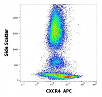
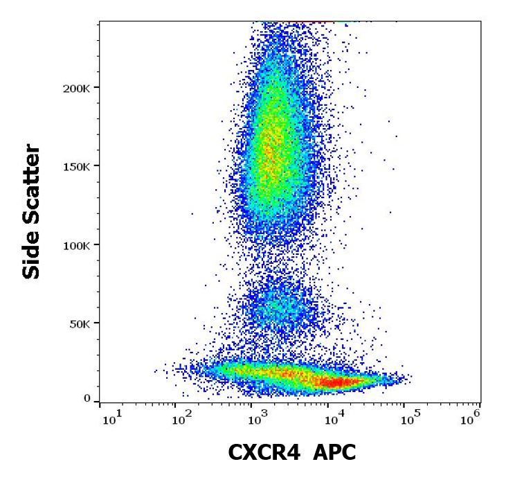
ARG42303 anti-CXCR4 antibody [12G5] (APC) FACS image
Flow Cytometry: Human peripheral whole blood stained with ARG42303 anti-CXCR4 antibody [12G5] (APC) at 10 µl / 100 µl of peripheral whole blood.
-

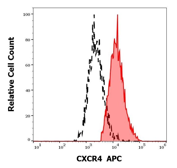
ARG42303 anti-CXCR4 antibody [12G5] (APC) FACS image
Flow Cytometry: Separation of Human CD184 positive lymphocytes (red-filled) from monocytes (black-dashed). Human peripheral whole blood stained with ARG42303 anti-CXCR4 antibody [12G5] (APC) at 10 µl / 100 µl of peripheral whole blood.