ARG23669
anti-CSF1R antibody [ROS-AV170]
anti-CSF1R antibody [ROS-AV170] for Flow cytometry,ICC/IF,IHC-Frozen sections and Chicken
Overview
| Product Description | Mouse Monoclonal antibody [ROS-AV170] recognizes CSF1R |
|---|---|
| Tested Reactivity | Chk |
| Species Does Not React With | Ms, Rat, Duck, Goose, Turkey |
| Tested Application | FACS, ICC/IF, IHC-Fr |
| Host | Mouse |
| Clonality | Monoclonal |
| Clone | ROS-AV170 |
| Isotype | IgG1 |
| Target Name | CSF1R |
| Antigen Species | Chicken |
| Immunogen | Purified ChCSF1R-Fc fusion protein. |
| Conjugation | Un-conjugated |
| Alternate Names | CSF-1 receptor; CSF-1-R; FIM2; Macrophage colony-stimulating factor 1 receptor; Proto-oncogene c-Fms; CD115; CD antigen CD115; M-CSF-R; FMS; CSFR; C-FMS; EC 2.7.10.1; CSF-1R; HDLS |
Application Instructions
| Application Suggestion |
|
||||||||
|---|---|---|---|---|---|---|---|---|---|
| Application Note | FACS: Use 10 µl of the suggested working dilution to label 10^6 cells in 100 µl. * The dilutions indicate recommended starting dilutions and the optimal dilutions or concentrations should be determined by the scientist. |
Properties
| Form | Liquid |
|---|---|
| Purification | Purification with Protein A. |
| Buffer | PBS and 0.09% Sodium azide. |
| Preservative | 0.09% Sodium azide |
| Concentration | 1 mg/ml |
| Storage Instruction | For continuous use, store undiluted antibody at 2-8°C for up to a week. For long-term storage, aliquot and store at -20°C or below. Storage in frost free freezers is not recommended. Avoid repeated freeze/thaw cycles. Suggest spin the vial prior to opening. The antibody solution should be gently mixed before use. |
| Note | For laboratory research only, not for drug, diagnostic or other use. |
Bioinformation
| Gene Symbol | CSF1R |
|---|---|
| Gene Full Name | colony stimulating factor 1 receptor |
| Background | The protein encoded by this gene is the receptor for colony stimulating factor 1, a cytokine which controls the production, differentiation, and function of macrophages. This receptor mediates most if not all of the biological effects of this cytokine. Ligand binding activates the receptor kinase through a process of oligomerization and transphosphorylation. The encoded protein is a tyrosine kinase transmembrane receptor and member of the CSF1/PDGF receptor family of tyrosine-protein kinases. Mutations in this gene have been associated with a predisposition to myeloid malignancy. The first intron of this gene contains a transcriptionally inactive ribosomal protein L7 processed pseudogene oriented in the opposite direction. Alternative splicing results in multiple transcript variants. [provided by RefSeq, Dec 2013] |
| Function | Tyrosine-protein kinase that acts as cell-surface receptor for CSF1 and IL34 and plays an essential role in the regulation of survival, proliferation and differentiation of hematopoietic precursor cells, especially mononuclear phagocytes, such as macrophages and monocytes. Promotes the release of proinflammatory chemokines in response to IL34 and CSF1, and thereby plays an important role in innate immunity and in inflammatory processes. Plays an important role in the regulation of osteoclast proliferation and differentiation, the regulation of bone resorption, and is required for normal bone and tooth development. Required for normal male and female fertility, and for normal development of milk ducts and acinar structures in the mammary gland during pregnancy. Promotes reorganization of the actin cytoskeleton, regulates formation of membrane ruffles, cell adhesion and cell migration, and promotes cancer cell invasion. Activates several signaling pathways in response to ligand binding. Phosphorylates PIK3R1, PLCG2, GRB2, SLA2 and CBL. Activation of PLCG2 leads to the production of the cellular signaling molecules diacylglycerol and inositol 1,4,5-trisphosphate, that then lead to the activation of protein kinase C family members, especially PRKCD. Phosphorylation of PIK3R1, the regulatory subunit of phosphatidylinositol 3-kinase, leads to activation of the AKT1 signaling pathway. Activated CSF1R also mediates activation of the MAP kinases MAPK1/ERK2 and/or MAPK3/ERK1, and of the SRC family kinases SRC, FYN and YES1. Activated CSF1R transmits signals both via proteins that directly interact with phosphorylated tyrosine residues in its intracellular domain, or via adapter proteins, such as GRB2. Promotes activation of STAT family members STAT3, STAT5A and/or STAT5B. Promotes tyrosine phosphorylation of SHC1 and INPP5D/SHIP-1. Receptor signaling is down-regulated by protein phosphatases, such as INPP5D/SHIP-1, that dephosphorylate the receptor and its downstream effectors, and by rapid internalization of the activated receptor. [UniProt] |
| Calculated MW | 108 kDa |
| PTM | Autophosphorylated in response to CSF1 or IL34 binding. Phosphorylation at Tyr-561 is important for normal down-regulation of signaling by ubiquitination, internalization and degradation. Phosphorylation at Tyr-561 and Tyr-809 is important for interaction with SRC family members, including FYN, YES1 and SRC, and for subsequent activation of these protein kinases. Phosphorylation at Tyr-699 and Tyr-923 is important for interaction with GRB2. Phosphorylation at Tyr-723 is important for interaction with PIK3R1. Phosphorylation at Tyr-708 is important for normal receptor degradation. Phosphorylation at Tyr-723 and Tyr-809 is important for interaction with PLCG2. Phosphorylation at Tyr-969 is important for interaction with CBL. Dephosphorylation by PTPN2 negatively regulates downstream signaling and macrophage differentiation. Ubiquitinated. Becomes rapidly polyubiquitinated after autophosphorylation, leading to its degradation. [UniProt] |
Images (4) Click the Picture to Zoom In
-
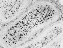
ARG23669 anti-CSF1R antibody [ROS-AV170] IHC-Fr image
Immunohistochemistry: Quail, Cortunix cortunix, bursa of Fabricius. Cryostat sections were cut from 4 week old Quail bursa of Fabricius. ARG23669 anti-CSF1R antibody [ROS-AV170] specifically stains the bursal secretory dendritic cells (BSDC) located in the medulla and the macrophages of the interfollicular connective tissue.
-
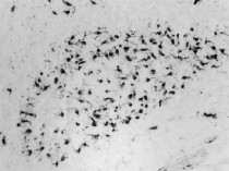
ARG23669 anti-CSF1R antibody [ROS-AV170] IHC-Fr image
Immunohistochemistry: Guinea fowl, Agelastes meleagredisbursa of Fabricius. ARG23669 anti-CSF1R antibody [ROS-AV170] specifically stains the bursal secretory dendritic cells (BSDC) located in the medulla. The antibody also recognizes some CSF1R+ve macrophages in the interfollicular connective tissue.
-
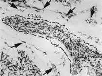
ARG23669 anti-CSF1R antibody [ROS-AV170] IHC-Fr image
Immunohistochemistry: Grey Patridge, Perdix perdix bursa of Fabricius. Cryostat sections cut from 4 week old partridge bursa of Fabricius. ARG23669 anti-CSF1R antibody [ROS-AV170] specifically recognized membrane expressed antigen on the bursal secretory dendritic cells (BSDC) in the medulla and by the macrophages (arrows) of the interfollicular connective tissue.
-
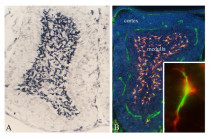
ARG23669 anti-CSF1R antibody [ROS-AV170] IHC-Fr image
Immunohistochemistry: Staining of Chicken bursa of Fabricius. Cryostat sections of 6 week old chicken bursa of Fabricius. A) ARG23669 anti-CSF1R antibody [ROS-AV170] identifies strong membrane-bound immunoreaction on the cell surface of the bursal secretory dendritic cells (BSDC). B) Double immunofluorescence staining with Mouse anti-Vimentin and ARG23669 (red).


