ARG58420
anti-CPB1 / Carboxypeptidase B antibody
anti-CPB1 / Carboxypeptidase B antibody for Flow cytometry,ICC/IF,IHC-Formalin-fixed paraffin-embedded sections,Western blot and Human
Overview
| Product Description | Rabbit Polyclonal antibody recognizes CPB1 / Carboxypeptidase B |
|---|---|
| Tested Reactivity | Hu |
| Tested Application | FACS, ICC/IF, IHC-P, WB |
| Host | Rabbit |
| Clonality | Polyclonal |
| Isotype | IgG |
| Target Name | CPB1 / Carboxypeptidase B |
| Antigen Species | Human |
| Immunogen | KLH-conjugated synthetic peptide corresponding to aa. 9-37 (N-terminus) of Human CPB1. |
| Conjugation | Un-conjugated |
| Alternate Names | Pancreas-specific protein; EC 3.4.17.2; Carboxypeptidase B; CPB; PASP; PCPB |
Application Instructions
| Application Suggestion |
|
||||||||||
|---|---|---|---|---|---|---|---|---|---|---|---|
| Application Note | * The dilutions indicate recommended starting dilutions and the optimal dilutions or concentrations should be determined by the scientist. | ||||||||||
| Positive Control | K562 |
Properties
| Form | Liquid |
|---|---|
| Purification | Purification with Protein A and immunogen peptide. |
| Buffer | PBS and 0.09% (W/V) Sodium azide. |
| Preservative | 0.09% (W/V) Sodium azide. |
| Storage Instruction | For continuous use, store undiluted antibody at 2-8°C for up to a week. For long-term storage, aliquot and store at -20°C or below. Storage in frost free freezers is not recommended. Avoid repeated freeze/thaw cycles. Suggest spin the vial prior to opening. The antibody solution should be gently mixed before use. |
| Note | For laboratory research only, not for drug, diagnostic or other use. |
Bioinformation
| Database Links | |
|---|---|
| Gene Symbol | CPB1 |
| Gene Full Name | carboxypeptidase B1 (tissue) |
| Background | Three different procarboxypeptidases A and two different procarboxypeptidases B have been isolated. The B1 and B2 forms differ from each other mainly in isoelectric point. Carboxypeptidase B1 is a highly tissue-specific protein and is a useful serum marker for acute pancreatitis and dysfunction of pancreatic transplants. It is not elevated in pancreatic carcinoma. [provided by RefSeq, Jul 2008] |
| Cellular Localization | Secreted. [UniProt] |
| Calculated MW | 47 kDa |
Images (4) Click the Picture to Zoom In
-
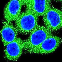
ARG58420 anti-CPB1 / Carboxypeptidase B antibody ICC/IF image
Immunofluorescence: HeLa cells stained with ARG58420 anti-CPB1 / Carboxypeptidase B antibody (green). DAPI (blue) for nuclear staining.
-
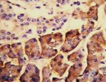
ARG58420 anti-CPB1 / Carboxypeptidase B antibody IHC-P image
Immunohistochemistry: Formalin-fixed and paraffin-embedded Human pancreas tissue stained with ARG58420 anti-CPB1 / Carboxypeptidase B antibody.
-
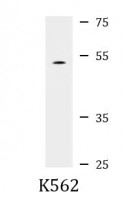
ARG58420 anti-CPB1 / Carboxypeptidase B antibody WB image
Western blot: 35 µg of K562 cell lysate stained with ARG58420 anti-CPB1 / Carboxypeptidase B antibody.
-

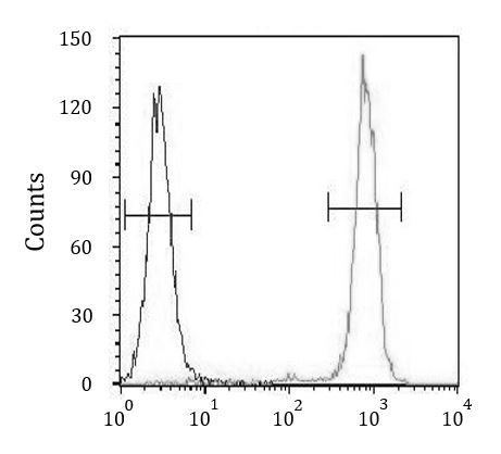
ARG58420 anti-CPB1 / Carboxypeptidase B antibody FACS image
Flow Cytometry: HeLa cells stained with ARG58420 anti-CPB1 / Carboxypeptidase B antibody (right histogram) or without primary antibody as control (left histogram), followed by incubation with FITC labelled secondary antibody.