ARG45189
anti-COX8A antibody
anti-COX8A antibody for Flow cytometry,IHC-Formalin-fixed paraffin-embedded sections,Western blot and Mouse,Rat
Overview
| Product Description | Rabbit Polyclonal antibody recognizes COX8A |
|---|---|
| Tested Reactivity | Ms, Rat |
| Tested Application | FACS, IHC-P, WB |
| Host | Rabbit |
| Clonality | Polyclonal |
| Isotype | IgG |
| Target Name | COX8A |
| Antigen Species | Human |
| Immunogen | Synthetic peptide corresponding to C-terminal region of mouse COX8A. |
| Conjugation | Un-conjugated |
| Alternate Names | COX8A; Cytochrome C Oxidase Subunit 8A; COX8L; COX8-2; VIII-L; VIII; COX8; COX; Cytochrome C Oxidase Polypeptide VIII-Liver/Heart; Cytochrome C Oxidase Subunit VIIIA (Ubiquitous); Cytochrome C Oxidase Subunit 8A, Mitochondrial; Cytochrome C Oxidase Subunit VIII; Cytochrome C Oxidase Subunit 8-2; Cytochrome C Oxidase Subunit 8A (Ubiquitous); MC4DN15 |
Application Instructions
| Application Suggestion |
|
||||||||
|---|---|---|---|---|---|---|---|---|---|
| Application Note | * The dilutions indicate recommended starting dilutions and the optimal dilutions or concentrations should be determined by the scientist. | ||||||||
| Observed Size | 8-10 kDa |
Properties
| Form | Liquid |
|---|---|
| Purification | Affinity purification with immunogen. |
| Buffer | 0.9% NaCl, 0.2% Na2HPO4, 0.01% Sodium azide and 4% Trehalose. |
| Preservative | 0.01% Sodium azide |
| Stabilizer | 4% Trehalose |
| Concentration | 0.5 mg/ml |
| Storage Instruction | For continuous use, store undiluted antibody at 2-8°C for up to a week. For long-term storage, aliquot and store at -20°C or below. Storage in frost free freezers is not recommended. Avoid repeated freeze/thaw cycles. Suggest spin the vial prior to opening. The antibody solution should be gently mixed before use. |
| Note | For laboratory research only, not for drug, diagnostic or other use. |
Bioinformation
| Gene Symbol | COX8A |
|---|---|
| Gene Full Name | Cytochrome C Oxidase Subunit 8A |
| Background | The protein encoded by this gene is the terminal enzyme of the respiratory chain, coupling the transfer of electrons from cytochrome c to molecular oxygen, with the concomitant production of a proton electrochemical gradient across the inner mitochondrial membrane. In addition to 3 mitochondrially encoded subunits, which perform the catalytic function, the eukaryotic enzyme contains nuclear-encoded smaller subunits, ranging in number from 4 in some organisms to 10 in mammals. It has been proposed that nuclear-encoded subunits may be involved in the modulation of the catalytic function. This gene encodes one of the nuclear-encoded subunits. [provided by RefSeq, Jul 2008] |
| Function | Component of the cytochrome c oxidase, the last enzyme in the mitochondrial electron transport chain which drives oxidative phosphorylation. The respiratory chain contains 3 multisubunit complexes succinate dehydrogenase (complex II, CII), ubiquinol-cytochrome c oxidoreductase (cytochrome b-c1 complex, complex III, CIII) and cytochrome c oxidase (complex IV, CIV), that cooperate to transfer electrons derived from NADH and succinate to molecular oxygen, creating an electrochemical gradient over the inner membrane that drives transmembrane transport and the ATP synthase. Cytochrome c oxidase is the component of the respiratory chain that catalyzes the reduction of oxygen to water. Electrons originating from reduced cytochrome c in the intermembrane space (IMS) are transferred via the dinuclear copper A center (CU(A)) of subunit 2 and heme A of subunit 1 to the active site in subunit 1, a binuclear center (BNC) formed by heme A3 and copper B (CU(B)). The BNC reduces molecular oxygen to 2 water molecules using 4 electrons from cytochrome c in the IMS and 4 protons from the mitochondrial matrix. [UniProt] |
| Cellular Localization | Mitochondrion inner membrane. [UniProt] |
| Calculated MW | 8 kDa |
| PTM | Ubl conjugation. [UniProt] |
Images (4) Click the Picture to Zoom In
-
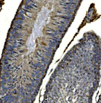
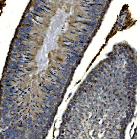
ARG45189 anti-COX8A antibody IHC-P image
Immunohistochemistry: Rat testis stained with ARG45189 anti-COX8A antibody at 2 μg/ml dilution.
-
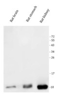
ARG45189 anti-COX8A antibody WB image
Western blot: Rat brain, rat stomach, and rat kidney stained with ARG45189 anti-COX8A antibody at 0.5 μg/ml dilution.
-

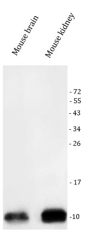
ARG45189 anti-COX8A antibody WB image
Western blot: Mouse brain and mouse kidney stained with ARG45189 anti-COX8A antibody at 0.5 μg/ml dilution.
-
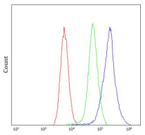
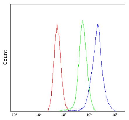
ARG45189 anti-COX8A antibody FACS image
Flow Cytometry: Mouse spleen stained with ARG45189 anti-COX8A antibody at 1 µg/10^6 cells dilution.