ARG63562
anti-COX2 antibody
anti-COX2 antibody for Flow cytometry,ICC/IF,Western blot and Human,Mouse
Inflammation Study antibody
Overview
| Product Description | Goat Polyclonal antibody recognizes COX2 |
|---|---|
| Tested Reactivity | Hu, Ms |
| Predict Reactivity | Cow, Dog, Pig |
| Tested Application | FACS, ICC/IF, WB |
| Host | Goat |
| Clonality | Polyclonal |
| Isotype | IgG |
| Target Name | COX2 |
| Antigen Species | Human |
| Immunogen | C-NPTVLLKERSTEL |
| Conjugation | Un-conjugated |
| Alternate Names | PHS II; Prostaglandin H2 synthase 2; PHS-2; Cyclooxygenase-2; PGHS-2; COX2; PGG/HS; COX-2; GRIPGHS; hCox-2; PGH synthase 2; Prostaglandin G/H synthase 2; Prostaglandin-endoperoxide synthase 2; EC 1.14.99.1 |
Application Instructions
| Application Suggestion |
|
||||||||
|---|---|---|---|---|---|---|---|---|---|
| Application Note | WB: Recommend incubate at RT for 1h. * The dilutions indicate recommended starting dilutions and the optimal dilutions or concentrations should be determined by the scientist. |
Properties
| Form | Liquid |
|---|---|
| Purification | Purified from goat serum by antigen affinity chromatography. |
| Buffer | Tris saline (pH 7.3), 0.02% Sodium azide and 0.5% BSA. |
| Preservative | 0.02% Sodium azide |
| Stabilizer | 0.5% BSA |
| Concentration | 0.5 mg/ml |
| Storage Instruction | For continuous use, store undiluted antibody at 2-8°C for up to a week. For long-term storage, aliquot and store at -20°C or below. Storage in frost free freezers is not recommended. Avoid repeated freeze/thaw cycles. Suggest spin the vial prior to opening. The antibody solution should be gently mixed before use. |
| Note | For laboratory research only, not for drug, diagnostic or other use. |
Bioinformation
| Database Links | |
|---|---|
| Background | COX2: Prostaglandin-endoperoxide synthase (PTGS), also known as cyclooxygenase, is the key enzyme in prostaglandin biosynthesis, and acts both as a dioxygenase and as a peroxidase. There are two isozymes of PTGS: a constitutive PTGS1 and an inducible PTGS2, which differ in their regulation of expression and tissue distribution. This gene encodes the inducible isozyme. It is regulated by specific stimulatory events, suggesting that it is responsible for the prostanoid biosynthesis involved in inflammation and mitogenesis. [provided by RefSeq, Feb 2009] |
| Function | COX2 converts arachidonate to prostaglandin H2 (PGH2), a committed step in prostanoid synthesis (PubMed:26859324, PubMed:27226593). Constitutively expressed in some tissues in physiological conditions, such as the endothelium, kidney and brain, and in pathological conditions, such as in cancer. PTGS2 is responsible for production of inflammatory prostaglandins. Up-regulation of PTGS2 is also associated with increased cell adhesion, phenotypic changes, resistance to apoptosis and tumor angiogenesis. In cancer cells, PTGS2 is a key step in the production of prostaglandin E2 (PGE2), which plays important roles in modulating motility, proliferation and resistance to apoptosis. During neuroinflammation, plays a role in neuronal secretion of specialized preresolving mediators (SPMs), especially 15-R-lipoxin A4, that regulates phagocytic microglia. [UniProt] |
| Highlight | Related products: COX2 antibodies; COX2 Duos / Panels; Anti-Goat IgG secondary antibodies; Related news: Exploring Antiviral Immune Response |
| Research Area | Inflammation Study antibody |
| Calculated MW | 69 kDa |
| PTM | S-nitrosylation by NOS2 (iNOS) activates enzyme activity. S-nitrosylation may take place on different Cys residues in addition to Cys-526. |
Images (5) Click the Picture to Zoom In
-
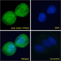
ARG63562 anti-COX2 antibody ICC/IF image
Immunofluorescence: Paraformaldehyde fixed HepG2 cells permeabilized with 0.15% Triton. Cells were stained with ARG63562 anti-COX2 antibody (green) at 10 µg/ml dilution for 1 hour. DAPI (blue) for nuclear staining. Negative control: Unimmunized goat IgG (green) at 10 µg/ml dilution.
-
ARG63562 anti-COX2 antibody WB image
Western blot: 35 µg of H460 cell lysate (in RIPA buffer) stained with ARG63562 anti-COX2 antibody at 0.5 µg/ml dilution and incubated at RT for 1 hour.
-
ARG63562 anti-COX2 antibody WB image
Western blot: 35 µg of A549 (A) and Daudi (B) cell lysates (in RIPA buffer) stained with ARG63562 anti-COX2 antibody at 0.1 µg/ml dilution and incubated at RT for 1 hour.
-
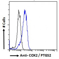
ARG63562 anti-COX2 antibody FACS image
Flow Cytometry: Paraformaldehyde-fixed HeLa cells permeabilized with 0.5% Triton. Cells were stained with ARG63562 anti-COX2 antibody (blue line) at 10 µg/ml dilution for 1 hour, followed by incubation with Alexa FluorR 488 labelled secondary antibody. IgG control: Unimmunized goat IgG (black line).
-
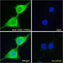
ARG63562 anti-COX2 antibody ICC/IF image
Immunofluorescence: Paraformaldehyde fixed NIH/3T3 cells permeabilized with 0.15% Triton. Cells were stained with ARG63562 anti-COX2 antibody (green) at 10 µg/ml dilution for 1 hour. DAPI (blue) for nuclear staining. Negative control: Unimmunized goat IgG (green) at 10 µg/ml dilution.


