ARG45628
anti-COPE antibody
anti-COPE antibody for Flow cytometry,ICC/IF,IHC-Frozen sections,IHC-Formalin-fixed paraffin-embedded sections,Western blot and Human,Mouse,Rat
Overview
| Product Description | Rabbit Polyclonal antibody recognizes COPE |
|---|---|
| Tested Reactivity | Hu, Ms, Rat |
| Tested Application | FACS, ICC/IF, IHC-Fr, IHC-P, WB |
| Host | Rabbit |
| Clonality | Polyclonal |
| Isotype | IgG |
| Target Name | COPE |
| Antigen Species | Human |
| Immunogen | Recombinant protein containing to human COPE. |
| Conjugation | Un-conjugated |
| Alternate Names | COPE; coatomer protein complex, subunit epsilon; Epsilon-COP; Epsilon-coat protein; epsilon-COP; Coatomer subunit epsilon |
Application Instructions
| Application Suggestion |
|
||||||||||||
|---|---|---|---|---|---|---|---|---|---|---|---|---|---|
| Application Note | * The dilutions indicate recommended starting dilutions and the optimal dilutions or concentrations should be determined by the scientist. | ||||||||||||
| Observed Size | 34 kDa |
Properties
| Form | Liquid |
|---|---|
| Purification | Affinity purified |
| Buffer | 0.9% NaCl, 0.2% Na2HPO4, 0.05% Sodium azide and 4% Trehalose. |
| Preservative | 0.05% Sodium azide |
| Stabilizer | 4% Trehalose |
| Concentration | 0.5 mg/ml |
| Storage Instruction | For continuous use, store undiluted antibody at 2-8°C for up to a week. For long-term storage, aliquot and store at -20°C or below. Storage in frost free freezers is not recommended. Avoid repeated freeze/thaw cycles. Suggest spin the vial prior to opening. The antibody solution should be gently mixed before use. |
| Note | For laboratory research only, not for drug, diagnostic or other use. |
Bioinformation
| Database Links | |
|---|---|
| Gene Symbol | COPE |
| Gene Full Name | coatomer protein complex, subunit epsilon |
| Background | The product of this gene is an epsilon subunit of coatomer protein complex. Coatomer is a cytosolic protein complex that binds to dilysine motifs and reversibly associates with Golgi non-clathrin-coated vesicles. It is required for budding from Golgi membranes, and is essential for the retrograde Golgi-to-ER transport of dilysine-tagged proteins. Coatomer complex consists of at least the alpha, beta, beta', gamma, delta, epsilon and zeta subunits. Alternatively spliced transcript variants encoding different isoforms have been identified. [provided by RefSeq, Jul 2008] |
| Function | The coatomer is a cytosolic protein complex that binds to dilysine motifs and reversibly associates with Golgi non-clathrin-coated vesicles, which further mediate biosynthetic protein transport from the ER, via the Golgi up to the trans Golgi network. The coatomer complex is required for budding from Golgi membranes, and is essential for the retrograde Golgi-to-ER transport of dilysine-tagged proteins. In mammals, the coatomer can only be recruited by membranes associated with ADP-ribosylation factors (ARFs), which are small GTP-binding proteins; the complex also influences the Golgi structural integrity, as well as the processing, activity, and endocytic recycling of LDL receptors (By similarity). [UniProt] |
| Cellular Localization | Cytoplasm; Cytoplasmic vesicle; Golgi apparatus; Membrane. [UniProt] |
| Calculated MW | 34 kDa |
| PTM | Phosphorylated by PKA. Polyubiquitinated by RCHY1 in the presence of androgen, leading to proteasomal degradation. [UniProt] |
Images (8) Click the Picture to Zoom In
-
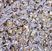
ARG45628 anti-COPE antibody IHC-P image
Immunohistochemistry: Human mammary cancer stained with ARG45628 anti-COPE antibody at 2 μg/ml dilution.
-

ARG45628 anti-COPE antibody ICC/IF image
Immunofluorescence: MCF-7 stained with ARG45628 anti-COPE antibody at 2 μg/ml dilution.
-

ARG45628 anti-COPE antibody WB image
Western blot: MCF-7 stained with ARG45628 anti-COPE antibody at 0.5 μg/ml dilution.
-

ARG45628 anti-COPE antibody FACS image
Flow Cytometry: HepG2 stained with ARG45628 anti-COPE antibody at 1 µg/10^6 cells dilution.
-
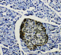
ARG45628 anti-COPE antibody IHC-P image
Immunohistochemistry: Rat pancreas stained with ARG45628 anti-COPE antibody at 2 μg/ml dilution.
-

ARG45628 anti-COPE antibody WB image
Western blot: Rat stomach stained with ARG45628 anti-COPE antibody at 0.5 μg/ml dilution.
-
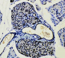
ARG45628 anti-COPE antibody IHC-P image
Immunohistochemistry: Mouse pancreas stained with ARG45628 anti-COPE antibody at 2 μg/ml dilution.
-

ARG45628 anti-COPE antibody WB image
Western blot: Mouse stomach stained with ARG45628 anti-COPE antibody at 0.5 μg/ml dilution.


