ARG66732
anti-COL18A1 antibody
anti-COL18A1 antibody for ICC/IF,IHC-Formalin-fixed paraffin-embedded sections,Western blot and Human
Overview
| Product Description | Rabbit Polyclonal antibody recognizes COL18A1 |
|---|---|
| Tested Reactivity | Hu |
| Tested Application | ICC/IF, IHC-P, WB |
| Host | Rabbit |
| Clonality | Polyclonal |
| Isotype | IgG |
| Target Name | COL18A1 |
| Antigen Species | Human |
| Immunogen | KLH-conjugated synthetic peptide within the center region of Human COL18A1. |
| Conjugation | Un-conjugated |
| Alternate Names | KS; KNO; KNO1; Collagen alpha-1(XVIII) chain |
Application Instructions
| Application Suggestion |
|
||||||||
|---|---|---|---|---|---|---|---|---|---|
| Application Note | IHC-P: Antigen Retrieval: Heat mediation was performed in Sodium citrate buffer (pH 6.0). * The dilutions indicate recommended starting dilutions and the optimal dilutions or concentrations should be determined by the scientist. |
||||||||
| Positive Control | MCF7 | ||||||||
| Observed Size | ~ 170 kDa |
Properties
| Form | Liquid |
|---|---|
| Purification | Affinity purification with immunogen. |
| Buffer | 0.42% Potassium phosphate (pH 7.3), 0.87% NaCl, 0.01% Sodium azide and 30% Glycerol. |
| Preservative | 0.01% Sodium azide |
| Stabilizer | 30% Glycerol |
| Storage Instruction | For continuous use, store undiluted antibody at 2-8°C for up to a week. For long-term storage, aliquot and store at -20°C. Storage in frost free freezers is not recommended. Avoid repeated freeze/thaw cycles. Suggest spin the vial prior to opening. The antibody solution should be gently mixed before use. |
| Note | For laboratory research only, not for drug, diagnostic or other use. |
Bioinformation
| Database Links | |
|---|---|
| Gene Symbol | COL18A1 |
| Gene Full Name | collagen, type XVIII, alpha 1 |
| Background | This gene encodes the alpha chain of type XVIII collagen. This collagen is one of the multiplexins, extracellular matrix proteins that contain multiple triple-helix domains (collagenous domains) interrupted by non-collagenous domains. A long isoform of the protein has an N-terminal domain that is homologous to the extracellular part of frizzled receptors. Proteolytic processing at several endogenous cleavage sites in the C-terminal domain results in production of endostatin, a potent antiangiogenic protein that is able to inhibit angiogenesis and tumor growth. Mutations in this gene are associated with Knobloch syndrome. The main features of this syndrome involve retinal abnormalities, so type XVIII collagen may play an important role in retinal structure and in neural tube closure. Alternative splicing results in multiple transcript variants. [provided by RefSeq, Dec 2014] |
| Function | Probably plays a major role in determining the retinal structure as well as in the closure of the neural tube. [Non-collagenous domain 1]: May regulate extracellular matrix-dependent motility and morphogenesis of endothelial and non-endothelial cells; the function requires homotrimerization and implicates MAPK signaling. Endostatin: Potently inhibits endothelial cell proliferation and angiogenesis (PubMed:9459295). May inhibit angiogenesis by binding to the heparan sulfate proteoglycans involved in growth factor signaling (By similarity). Inhibits VEGFA-induced endothelial cell proliferation and migration. Seems to inhibit VEGFA-mediated signaling by blocking the interaction of VEGFA to its receptor KDR/VEGFR2. Modulates endothelial cell migration in an integrin-dependent manner implicating integrin ITGA5:ITGB1 and to a lesser extent ITGAV:ITGB3 and ITGAV:ITGB5 (By similarity). May negatively regulate the activity of homotrimeric non-collagenous domain 1 (PubMed:11257123). [UniProt] |
| Cellular Localization | Secreted, extracellular space, extracellular matrix. Secreted, extracellular space, extracellular matrix, basement membrane. Non-collagenous domain 1: Secreted, extracellular space, extracellular matrix, basement membrane. Secreted. Endostatin: Secreted. Secreted, extracellular space, extracellular matrix, basement membrane. [UniProt] |
| Calculated MW | 178 kDa |
| PTM | Prolines at the third position of the tripeptide repeating unit (G-X-Y) of the triple-helical regions are hydroxylated. [UniProt] |
Images (3) Click the Picture to Zoom In
-
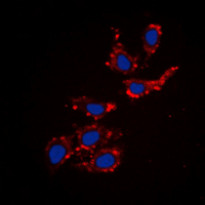
ARG66732 anti-COL18A1 antibody ICC/IF image
Immunofluorescence: Formalin-fixed MCF7 cells were permeabilized with 0.1% Triton X-100 in TBS for 5-10 minutes and blocked with 3% BSA-PBS for 30 minutes at room temperature. Cells were stained with ARG66732 anti-COL18A1 antibody (red) in 3% BSA-PBS and incubated overnight at 4°C. Cells were washed with PBST and incubated with a DyLight 594-conjugated secondary antibody in PBS at room temperature in the dark. DAPI (blue) for nuclear staining.
-
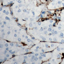
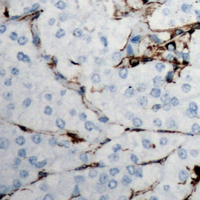
ARG66732 anti-COL18A1 antibody IHC-P image
Immunohistochemistry: Formalin-fixed and paraffin-embedded Human breast cancer tissue section. Antigen Retrieval: Heat mediation was performed in Sodium citrate buffer (pH 6.0). The section was stained with ARG66732 anti-COL18A1 antibody at room temperature. The section was counterstained with haematoxylin and mounted with DPX.
-
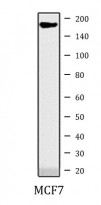
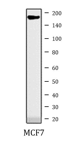
ARG66732 anti-COL18A1 antibody WB image
Western blot: MCF7 whole cell lysate stained with ARG66732 anti-COL18A1 antibody.