ARG64725
anti-COG7 antibody
anti-COG7 antibody for IHC-Formalin-fixed paraffin-embedded sections,Western blot and Human
Signaling Transduction antibody
Overview
| Product Description | Goat Polyclonal antibody recognizes COG7 |
|---|---|
| Tested Reactivity | Hu |
| Tested Application | IHC-P, WB |
| Host | Goat |
| Clonality | Polyclonal |
| Isotype | IgG |
| Target Name | COG7 |
| Antigen Species | Human |
| Immunogen | C-KTRPEDYRQVSK |
| Conjugation | Un-conjugated |
| Alternate Names | Conserved oligomeric Golgi complex subunit 7; Component of oligomeric Golgi complex 7; CDG2E; COG complex subunit 7 |
Application Instructions
| Application Suggestion |
|
||||||
|---|---|---|---|---|---|---|---|
| Application Note | WB: Recommend incubate at RT for 1h. IHC-P: Antigen Retrieval: Steam tissue section in Citrate buffer (pH 6.0). * The dilutions indicate recommended starting dilutions and the optimal dilutions or concentrations should be determined by the scientist. |
Properties
| Form | Liquid |
|---|---|
| Purification | Purified from goat serum by antigen affinity chromatography. |
| Buffer | Tris saline (pH 7.3), 0.02% Sodium azide and 0.5% BSA. |
| Preservative | 0.02% Sodium azide |
| Stabilizer | 0.5% BSA |
| Concentration | 0.5 mg/ml |
| Storage Instruction | For continuous use, store undiluted antibody at 2-8°C for up to a week. For long-term storage, aliquot and store at -20°C or below. Storage in frost free freezers is not recommended. Avoid repeated freeze/thaw cycles. Suggest spin the vial prior to opening. The antibody solution should be gently mixed before use. |
| Note | For laboratory research only, not for drug, diagnostic or other use. |
Bioinformation
| Database Links |
Swiss-port # P83436 Human Conserved oligomeric Golgi complex subunit 7 |
|---|---|
| Background | The protein encoded by this gene resides in the golgi, and constitutes one of the 8 subunits of the conserved oligomeric Golgi (COG) complex, which is required for normal golgi morphology and localization. Mutations in this gene are associated with the congenital disorder of glycosylation type IIe.[provided by RefSeq, May 2010] |
| Research Area | Signaling Transduction antibody |
| Calculated MW | 86 kDa |
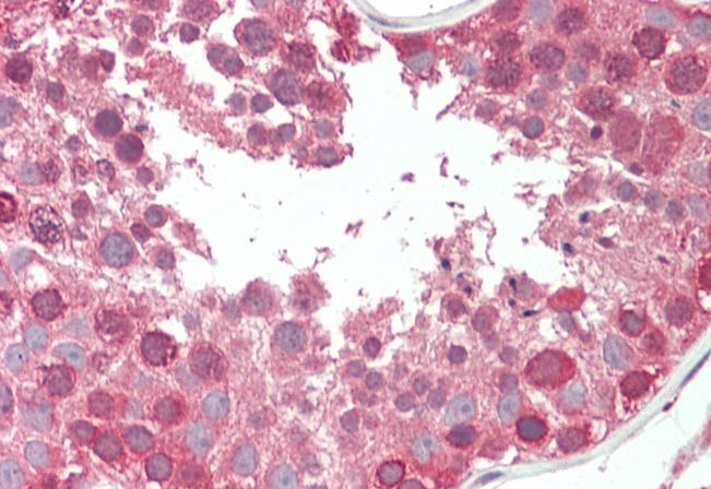
Images (2) Click the Picture to Zoom In
-
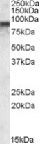
ARG64725 anti-COG7 antibody WB image
Western Blot: MOLT4 lysate (35 µg protein in RIPA buffer) stained with ARG64725 anti-COG7 antibody at 0.03 µg/ml dilution.
-

ARG64725 anti-COG7 antibody IHC-P image
Immunohistochemistry: Paraffin-embedded Human testis tissue. Antigen Retrieval: Steam tissue section in Citrate buffer (pH 6.0). The tissue section was stained with ARG64725 anti-COG7 antibody at 5 µg/ml dilution followed by AP-staining.