ARG10754
anti-CNPase antibody
anti-CNPase antibody for ICC/IF,IHC-Frozen sections,Western blot and Human,Mouse,Rat,Cow
Overview
| Product Description | Chicken Polyclonal antibody to CNP |
|---|---|
| Tested Reactivity | Hu, Ms, Rat, Cow |
| Tested Application | ICC/IF, IHC-Fr, WB |
| Host | Chicken |
| Clonality | Polyclonal |
| Isotype | IgY |
| Target Name | CNPase |
| Antigen Species | Human |
| Immunogen | Full length Human recombinant protein |
| Conjugation | Un-conjugated |
| Alternate Names | CNPase; 2',3'-cyclic-nucleotide 3'-phosphodiesterase; CNP1; CNP; EC 3.1.4.37 |
Application Instructions
| Application Suggestion |
|
||||||||
|---|---|---|---|---|---|---|---|---|---|
| Application Note | * The dilutions indicate recommended starting dilutions and the optimal dilutions or concentrations should be determined by the scientist. |
Properties
| Form | Liquid |
|---|---|
| Buffer | PBS and 0.02% Sodium azide. |
| Preservative | 0.02% Sodium azide |
| Storage Instruction | For continuous use, store undiluted antibody at 2-8°C for up to a week. For long-term storage, aliquot and store at -20°C or below. Storage in frost free freezers is not recommended. Avoid repeated freeze/thaw cycles. Suggest spin the vial prior to opening. The antibody solution should be gently mixed before use. |
| Note | For laboratory research only, not for drug, diagnostic or other use. |
Bioinformation
| Database Links | |
|---|---|
| Gene Symbol | CNP |
| Gene Full Name | 2',3'-cyclic nucleotide 3' phosphodiesterase |
| Function | May participate in RNA metabolism in the myelinating cell, CNP is the third most abundant protein in central nervous system myelin. [UniProt] |
| Calculated MW | 48 kDa |
Images (4) Click the Picture to Zoom In
-
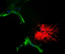
ARG10754 anti-CNPase antibody ICC/IF image
Immunocytochemistry: Rat mixed neuron-glial cell cultures stained with ARG10754 anti-CNPase antibody (red) and co-stained with a Monoclonal 5C10 against GFAP (green). The CNP antibody stains strongly in oligodendrocytes, whereas GFAP labels the intermediate filament in astrocytes. Blue is DNA staining showing the nuclei of these and numerous other cells, most of which are neurons and which do not express either CNP or GFAP.
-
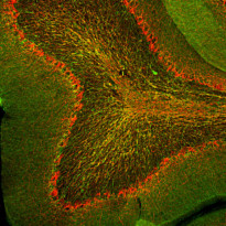
ARG10754 anti-CNPase antibody IHC-Fr image
Immunohistochemistry: Frozen section of Rat cerebellum stained with ARG10754 anti-CNPase antibody (green) at 1:2000 dilution and costained with ARG10761 anti-Neurofilament NF-H antibody (red) at 1:10000 dilution. (Sample preparation: Following transcardial perfusion of Rat with 4% paraformaldehyde, brain was post fixed for 24 hours, cut to 45 µM, and free-floating sections were stained with above antibodies.)
The CNPase antibody stains myelin and oligodendrocytes, cells that create the myelin sheath around axons. The NF-H antibody labels the heavily phosphorylated axonal forms of NF-H which are localized in large projection axons.
-
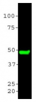
ARG10754 anti-CNPase antibody WB image
Western blot: Rat brain tissue homogenates stained with ARG10754 anti-CNPase antibody at 1:5000.
-
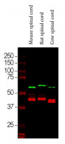
ARG10754 anti-CNPase antibody WB image
Western blot: Mouse spinal cord, Rat spinal cord and Cow spinal cord lysates stained with ARG10754 anti-CNPase antibody (red) at 1:5000 dilution. The blot was simultaneously stained with Mouse mAb to alpha Internexin (green) at 1:2000 dilution.


