ARG57010
anti-CNDP2 antibody [15E5]
anti-CNDP2 antibody [15E5] for ICC/IF,Western blot and Human
Overview
| Product Description | Mouse Monoclonal antibody [15E5] recognizes CNDP2 |
|---|---|
| Tested Reactivity | Hu |
| Tested Application | ICC/IF, WB |
| Host | Mouse |
| Clonality | Monoclonal |
| Clone | 15E5 |
| Isotype | IgG1, kappa |
| Target Name | CNDP2 |
| Antigen Species | Human |
| Immunogen | Recombinant fragment around aa. 1-475 of Human CNDP2. |
| Conjugation | Un-conjugated |
| Alternate Names | CN2; Peptidase A; PEPA; EC 3.4.13.18; CNDP dipeptidase 2; CPGL; HsT2298; HEL-S-13; Cytosolic non-specific dipeptidase; Glutamate carboxypeptidase-like protein 1 |
Application Instructions
| Application Suggestion |
|
||||||
|---|---|---|---|---|---|---|---|
| Application Note | * The dilutions indicate recommended starting dilutions and the optimal dilutions or concentrations should be determined by the scientist. |
Properties
| Form | Liquid |
|---|---|
| Purification | Purification with Protein G. |
| Buffer | PBS (pH 7.4), 0.02% Sodium azide and 10% Glycerol. |
| Preservative | 0.02% Sodium azide |
| Stabilizer | 10% Glycerol |
| Concentration | 1 mg/ml |
| Storage Instruction | For continuous use, store undiluted antibody at 2-8°C for up to a week. For long-term storage, aliquot and store at -20°C. Storage in frost free freezers is not recommended. Avoid repeated freeze/thaw cycles. Suggest spin the vial prior to opening. The antibody solution should be gently mixed before use. |
| Note | For laboratory research only, not for drug, diagnostic or other use. |
Bioinformation
| Database Links |
Swiss-port # Q96KP4 Human Cytosolic non-specific dipeptidase |
|---|---|
| Gene Symbol | CNDP2 |
| Gene Full Name | CNDP dipeptidase 2 (metallopeptidase M20 family) |
| Background | CNDP2, also known as tissue carnosinase and peptidase A (EC 3.4.13.18), is a nonspecific dipeptidase rather than a selective carnosinase (Teufel et al., 2003 [PubMed 12473676]).[supplied by OMIM, Mar 2008] |
| Function | Hydrolyzes a variety of dipeptides including L-carnosine but has a strong preference for Cys-Gly. Isoform 2 may be play a role as tumor suppressor in hepatocellular carcinoma (HCC) cells. [UniProt] |
| Calculated MW | 53 kDa |
Images (3) Click the Picture to Zoom In
-
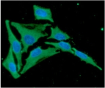
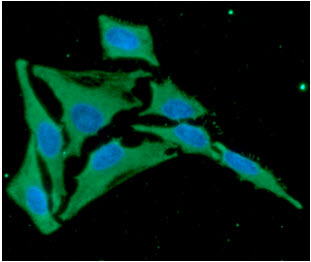
ARG57010 anti-CNDP2 antibody [15E5] ICC/IF image
Immunoflorescense: HeLa cell line stained with ARG57010 anti-CNDP2 antibody [15E5] at 1:100 (Green).
DAPI (Blue) for nucleus staining.
-
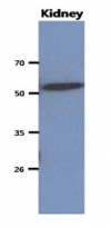
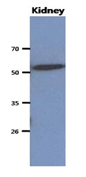
ARG57010 anti-CNDP2 antibody [15E5] WB image
Western blot: 40 µg of Mouse kidney stained with ARG57010 anti-CNDP2 antibody [15E5] at 1:1000.
-
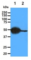
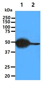
ARG57010 anti-CNDP2 antibody [15E5] WB image
Western blot: 40 µg of 1) Mouse Liver Tissue lysate, 2) HepG2 cell lysate stained with ARG57010 anti-CNDP2 antibody [15E5] at 1:1000.