ARG42067
anti-CLEC4M / DC-SIGNR / L-SIGN antibody
anti-CLEC4M / DC-SIGNR / L-SIGN antibody for IHC-Formalin-fixed paraffin-embedded sections,Western blot and Human
Overview
| Product Description | Rabbit Polyclonal antibody recognizes CLEC4M / DC-SIGNR / L-SIGN |
|---|---|
| Tested Reactivity | Hu |
| Predict Reactivity | Pig |
| Tested Application | IHC-P, WB |
| Host | Rabbit |
| Clonality | Polyclonal |
| Isotype | IgG |
| Target Name | CLEC4M / DC-SIGNR / L-SIGN |
| Antigen Species | Human |
| Immunogen | Synthetic peptide around the N-terminal region of Human CLEC4M. (within the following region: LVLQL LSFML LAGVL VAILV QVSKV PSSLS QEQSE QDAIY QNLTQ LKAAV) |
| Conjugation | Un-conjugated |
| Alternate Names | LSIGN; DC-SIGNR; CD209L; DCSIGNR; CD antigen CD299; Dendritic cell-specific ICAM-3-grabbing non-integrin 2; CD299; DC-SIGN-related protein; L-SIGN; Liver/lymph node-specific ICAM-3-grabbing non-integrin; HP10347; C-type lectin domain family 4 member M; DC-SIGN2; CD209 antigen-like protein 1 |
Application Instructions
| Predict Reactivity Note | Predicted Homology Based On Immunogen Sequence: Pig: 86% | ||||||
|---|---|---|---|---|---|---|---|
| Application Suggestion |
|
||||||
| Application Note | * The dilutions indicate recommended starting dilutions and the optimal dilutions or concentrations should be determined by the scientist. | ||||||
| Positive Control | HepG2 | ||||||
| Observed Size | ~ 57 kDa |
Properties
| Form | Liquid |
|---|---|
| Purification | Purification with Protein A. |
| Buffer | PBS, 0.09% (w/v) Sodium azide and 2% Sucrose. |
| Preservative | 0.09% (w/v) Sodium azide |
| Stabilizer | 2% Sucrose |
| Concentration | Batch dependent: 0.5 - 1 mg/ml |
| Storage Instruction | For continuous use, store undiluted antibody at 2-8°C for up to a week. For long-term storage, aliquot and store at -20°C or below. Storage in frost free freezers is not recommended. Avoid repeated freeze/thaw cycles. Suggest spin the vial prior to opening. The antibody solution should be gently mixed before use. |
| Note | For laboratory research only, not for drug, diagnostic or other use. |
Bioinformation
| Database Links |
Swiss-port # Q9H2X3 Human C-type lectin domain family 4 member M |
|---|---|
| Gene Symbol | CLEC4M |
| Gene Full Name | C-type lectin domain family 4, member M |
| Background | This gene encodes a transmembrane receptor and is often referred to as L-SIGN because of its expression in the endothelial cells of the lymph nodes and liver. The encoded protein is involved in the innate immune system and recognizes numerous evolutionarily divergent pathogens ranging from parasites to viruses, with a large impact on public health. The protein is organized into three distinct domains: an N-terminal transmembrane domain, a tandem-repeat neck domain and C-type lectin carbohydrate recognition domain. The extracellular region consisting of the C-type lectin and neck domains has a dual function as a pathogen recognition receptor and a cell adhesion receptor by binding carbohydrate ligands on the surface of microbes and endogenous cells. The neck region is important for homo-oligomerization which allows the receptor to bind multivalent ligands with high avidity. Variations in the number of 23 amino acid repeats in the neck domain of this protein are common and have a significant impact on ligand binding ability. This gene is closely related in terms of both sequence and function to a neighboring gene (GeneID 30835; often referred to as DC-SIGN or CD209). DC-SIGN and L-SIGN differ in their ligand-binding properties and distribution. Alternative splicing results in multiple variants. [provided by RefSeq, Feb 2009] |
| Function | Probable pathogen-recognition receptor involved in peripheral immune surveillance in liver. May mediate the endocytosis of pathogens which are subsequently degraded in lysosomal compartments. Is a receptor for ICAM3, probably by binding to mannose-like carbohydrates. (Microbial infection) Acts as an attachment receptor for Ebolavirus, Hepatitis C virus, HIV-1, Human coronavirus 229E, Human cytomegalovirus/HHV-5, Influenzavirus, SARS coronavirus, West-nile virus, Japanese encephalitis virus and Marburg virus glycoprotein. (Microbial infection) Recognition of M. bovis by dendritic cells may occur partially via this molecule. [UniProt] |
| Cellular Localization | Isoform 1, 2 and 3: Cell membrane; Single-pass type II membrane protein. Isoform 5, 6, 7 and 10: Secreted. [UniProt] |
| Calculated MW | 45 kDa |
Images (2) Click the Picture to Zoom In
-
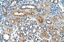
ARG42067 anti-CLEC4M / DC-SIGNR / L-SIGN antibody IHC-P image
Immunohistochemistry: Paraffin-embedded Human kidney tissue stained with ARG42067 anti-CLEC4M / DC-SIGNR / L-SIGN antibody.
-
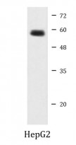
ARG42067 anti-CLEC4M / DC-SIGNR / L-SIGN antibody WB image
Western blot: HepG2 cell lysate stained with ARG42067 anti-CLEC4M / DC-SIGNR / L-SIGN antibody at 1.25 µg/ml dilution.


