ARG59049
anti-CIITA antibody
anti-CIITA antibody for IHC-Formalin-fixed paraffin-embedded sections,Western blot and Human,Mouse,Rat
Overview
| Product Description | Rabbit Polyclonal antibody recognizes CIITA |
|---|---|
| Tested Reactivity | Hu, Ms, Rat |
| Tested Application | IHC-P, WB |
| Host | Rabbit |
| Clonality | Polyclonal |
| Isotype | IgG |
| Target Name | CIITA |
| Antigen Species | Human |
| Immunogen | Recombinant protein corresponding to E945-R1130 of Human CIITA. |
| Conjugation | Un-conjugated |
| Alternate Names | MHC class II transactivator; NLRA; CIITA; C2TA; MHC2TA; EC 2.3.1.-; CIITAIV; EC 2.7.11.1 |
Application Instructions
| Application Suggestion |
|
||||||
|---|---|---|---|---|---|---|---|
| Application Note | IHC-P: Antigen Retrieval: By heat mediation. * The dilutions indicate recommended starting dilutions and the optimal dilutions or concentrations should be determined by the scientist. |
Properties
| Form | Liquid |
|---|---|
| Purification | Affinity purification with immunogen. |
| Buffer | 0.2% Na2HPO4, 0.9% NaCl, 0.05% Sodium azide and 5% BSA. |
| Preservative | 0.05% Sodium azide |
| Stabilizer | 5% BSA |
| Concentration | 0.5 mg/ml |
| Storage Instruction | For continuous use, store undiluted antibody at 2-8°C for up to a week. For long-term storage, aliquot and store at -20°C or below. Storage in frost free freezers is not recommended. Avoid repeated freeze/thaw cycles. Suggest spin the vial prior to opening. The antibody solution should be gently mixed before use. |
| Note | For laboratory research only, not for drug, diagnostic or other use. |
Bioinformation
| Database Links | |
|---|---|
| Gene Symbol | CIITA |
| Gene Full Name | class II, major histocompatibility complex, transactivator |
| Background | This gene encodes a protein with an acidic transcriptional activation domain, 4 LRRs (leucine-rich repeats) and a GTP binding domain. The protein is located in the nucleus and acts as a positive regulator of class II major histocompatibility complex gene transcription, and is referred to as the "master control factor" for the expression of these genes. The protein also binds GTP and uses GTP binding to facilitate its own transport into the nucleus. Once in the nucleus it does not bind DNA but rather uses an intrinsic acetyltransferase (AT) activity to act in a coactivator-like fashion. Mutations in this gene have been associated with bare lymphocyte syndrome type II (also known as hereditary MHC class II deficiency or HLA class II-deficient combined immunodeficiency), increased susceptibility to rheumatoid arthritis, multiple sclerosis, and possibly myocardial infarction. Several transcript variants encoding different isoforms have been found for this gene. [provided by RefSeq, Nov 2013] |
| Function | Essential for transcriptional activity of the HLA class II promoter; activation is via the proximal promoter. No DNA binding of in vitro translated CIITA was detected. May act in a coactivator-like fashion through protein-protein interactions by contacting factors binding to the proximal MHC class II promoter, to elements of the transcription machinery, or both. Alternatively it may activate HLA class II transcription by modifying proteins that bind to the MHC class II promoter. Also mediates enhanced MHC class I transcription; the promoter element requirements for CIITA-mediated transcription are distinct from those of constitutive MHC class I transcription, and CIITA can functionally replace TAF1 at these genes. Exhibits intrinsic GTP-stimulated acetyltransferase activity. Exhibits serine/threonine protein kinase activity: can phosphorylate the TFIID component TAF7, the RAP74 subunit of the general transcription factor TFIIF, histone H2B at 'Ser-37' and other histones (in vitro). [UniProt] |
| Cellular Localization | Nucleus. Nucleus, PML body. Recruited to PML body by PML. [UniProt] |
| Calculated MW | 124 kDa |
| PTM | Autophosphorylated, affecting interaction with TAF7. [UniProt] |
Images (4) Click the Picture to Zoom In
-

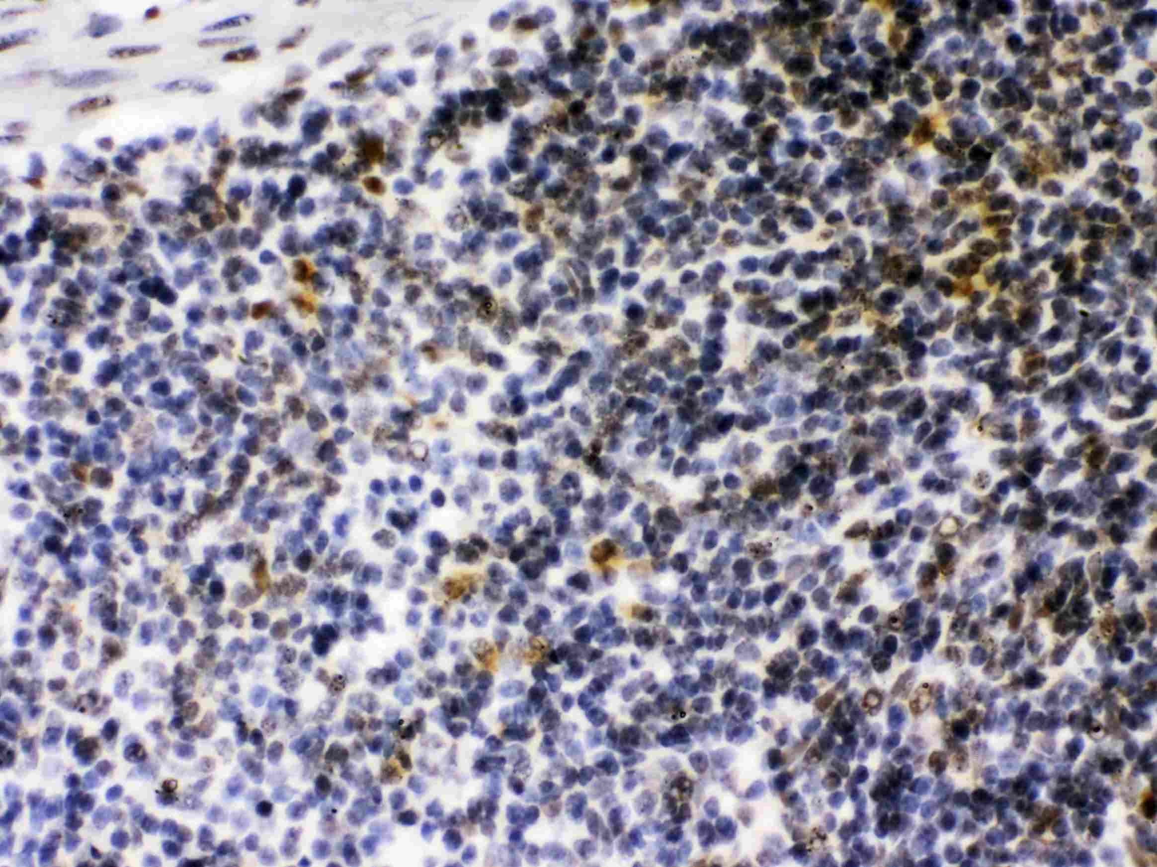
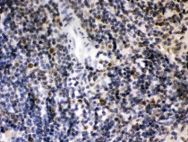
ARG59049 anti-CIITA antibody IHC-P image
Immunohistochemistry: Paraffin-embedded Mouse spleen tissue stained with ARG59049 anti-CIITA antibody at 1 µg/ml dilution.
-
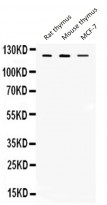
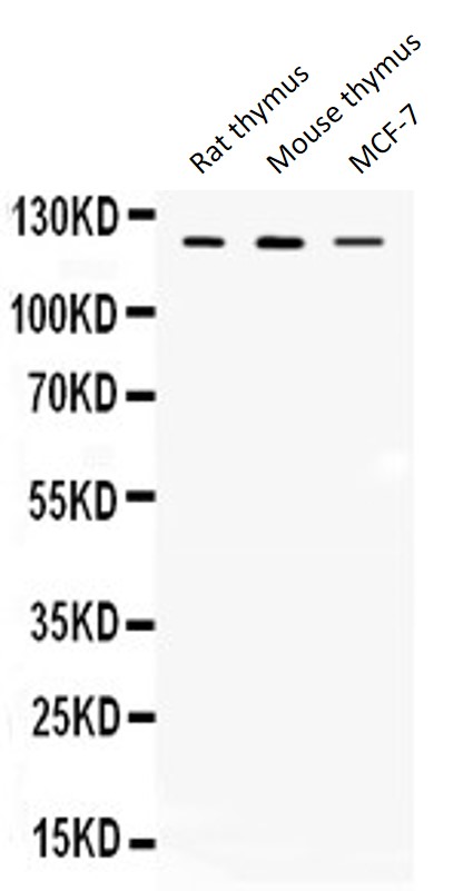
ARG59049 anti-CIITA antibody WB image
Western blot: Rat thymus, Mouse thymus and MCF-7 lysates stained with ARG59049 anti-CIITA antibody at 0.5 µg/ml dilution.
-
ARG59049 anti-CIITA antibody IHC-P image
Immunohistochemistry: Paraffin-embedded Rat spleen tissue stained with ARG59049 anti-CIITA antibody at 1 µg/ml dilution.
-
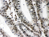
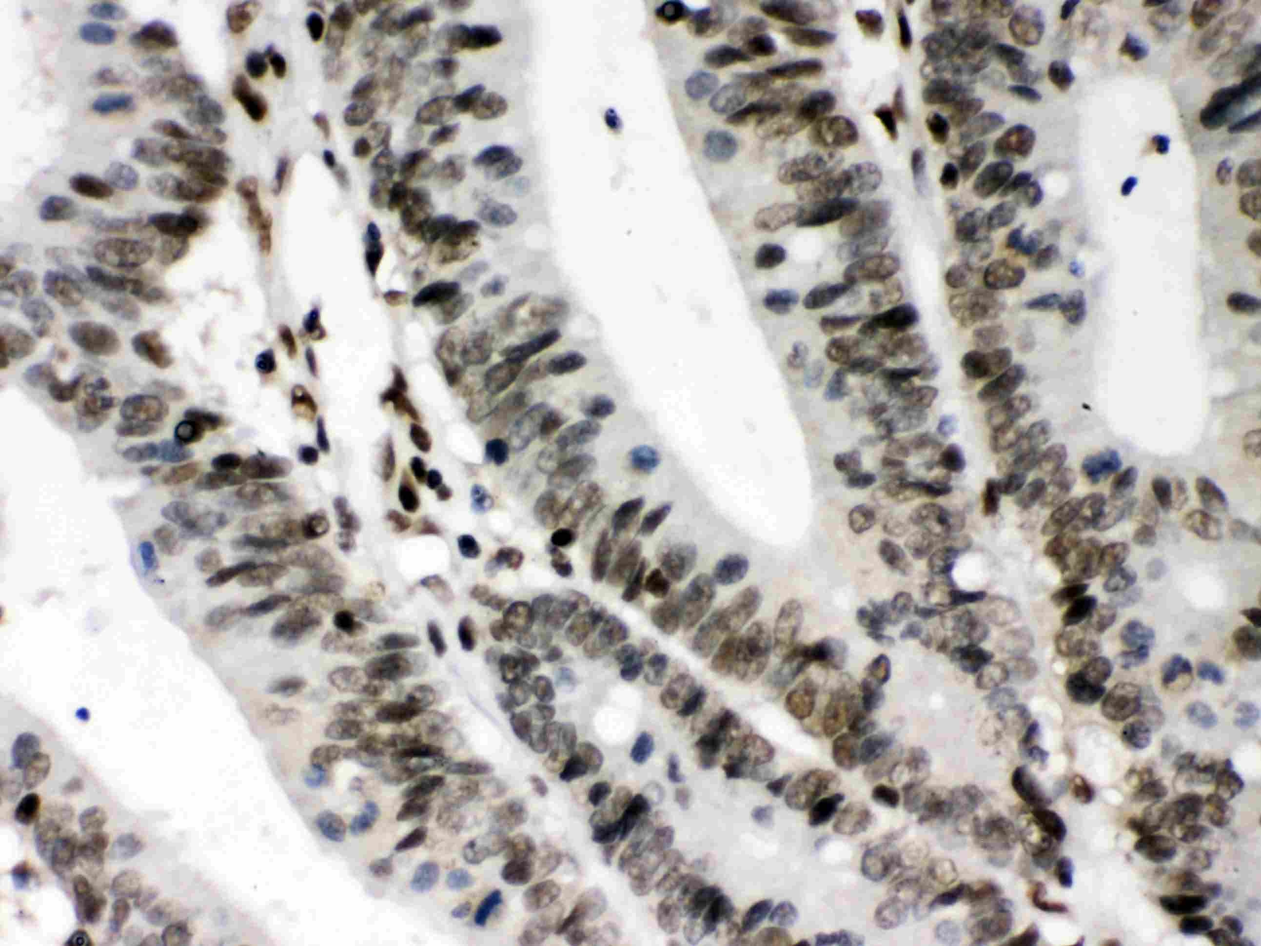
ARG59049 anti-CIITA antibody IHC-P image
Immunohistochemistry: Paraffin-embedded Human intestinal cancer tissue stained with ARG59049 anti-CIITA antibody at 1 µg/ml dilution.