ARG58428
anti-CHRNA5 antibody
anti-CHRNA5 antibody for Flow cytometry,IHC-Formalin-fixed paraffin-embedded sections,Western blot and Human,Mouse,Rat
Overview
| Product Description | Rabbit Polyclonal antibody recognizes CHRNA5 |
|---|---|
| Tested Reactivity | Hu, Ms, Rat |
| Tested Application | FACS, IHC-P, WB |
| Host | Rabbit |
| Clonality | Polyclonal |
| Isotype | IgG |
| Target Name | CHRNA5 |
| Antigen Species | Human |
| Immunogen | Synthetic peptide corresponding to a sequence at the N-terminus of Human CHRNA5 (44-76aa AKHEDSLLKDLFQDYERWVRPVEHLNDKIKIKF), different from the related Mouse sequence by five amino acids, and from the related Rat sequence by four amino acids. |
| Conjugation | Un-conjugated |
| Alternate Names | LNCR2; Neuronal acetylcholine receptor subunit alpha-5 |
Application Instructions
| Application Suggestion |
|
||||||||
|---|---|---|---|---|---|---|---|---|---|
| Application Note | IHC-P: Antigen Retrieval: Heat mediated was performed in Citrate buffer (pH 6.0) for 20 min. * The dilutions indicate recommended starting dilutions and the optimal dilutions or concentrations should be determined by the scientist. |
Properties
| Form | Liquid |
|---|---|
| Purification | Affinity purification with immunogen. |
| Buffer | 0.9% NaCl, 0.2% Na2HPO4, 0.05% Sodium azide and 5% BSA. |
| Preservative | 0.05% Sodium azide |
| Stabilizer | 5% BSA |
| Concentration | 0.5 mg/ml |
| Storage Instruction | For continuous use, store undiluted antibody at 2-8°C for up to a week. For long-term storage, aliquot and store at -20°C or below. Storage in frost free freezers is not recommended. Avoid repeated freeze/thaw cycles. Suggest spin the vial prior to opening. The antibody solution should be gently mixed before use. |
| Note | For laboratory research only, not for drug, diagnostic or other use. |
Bioinformation
| Database Links |
Swiss-port # P30532 Human Neuronal acetylcholine receptor subunit alpha-5 Swiss-port # Q2MKA5 Mouse Neuronal acetylcholine receptor subunit alpha-5 |
|---|---|
| Gene Symbol | CHRNA5 |
| Gene Full Name | cholinergic receptor, nicotinic, alpha 5 (neuronal) |
| Background | The protein encoded by this gene is a nicotinic acetylcholine receptor subunit and a member of a superfamily of ligand-gated ion channels that mediate fast signal transmission at synapses. These receptors are thought to be heteropentamers composed of separate but similar subunits. Defects in this gene have been linked to susceptibility to lung cancer type 2 (LNCR2).[provided by RefSeq, Jun 2010] |
| Function | After binding acetylcholine, the AChR responds by an extensive change in conformation that affects all subunits and leads to opening of an ion-conducting channel across the plasma membrane. [UniProt] |
| Cellular Localization | Cell junction, synapse, postsynaptic cell membrane; Multi-pass membrane protein. Cell membrane; Multi-pass membrane protein. [UniProt] |
| Calculated MW | 53 kDa |
Images (9) Click the Picture to Zoom In
-
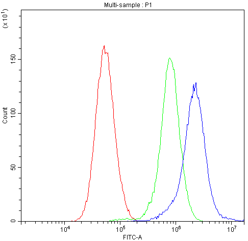
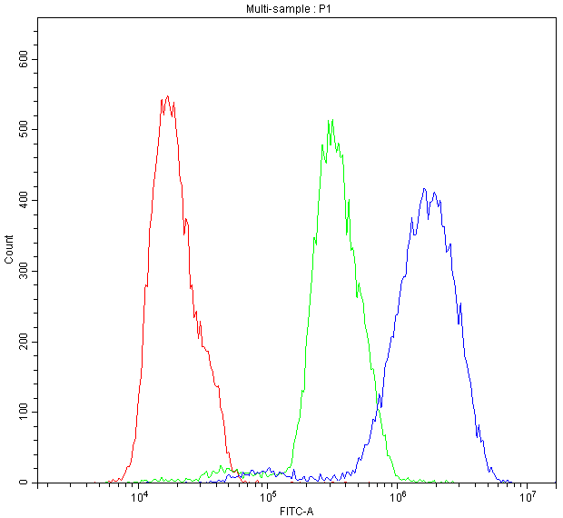
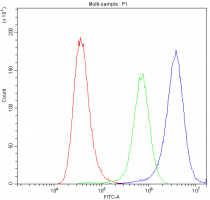
ARG58428 anti-CHRNA5 antibody FACS image
Flow Cytometry: U251 cells were blocked with 10% normal goat serum, and then stained with ARG58428 anti-CHRNA5 antibody (blue) at 1 µg/10^6 cells for 30 min at 20°C, followed by DyLight®488 labelled secondary antibody. Isotype control antibody (green) was rabbit IgG (1 µg/10^6 cells) used under the same conditions. Unlabelled sample (red) was also used as a control.
-
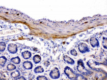
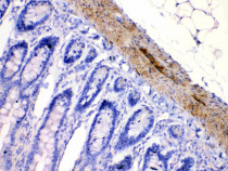
ARG58428 anti-CHRNA5 antibody IHC-P image
Immunohistochemistry: Paraffin-embedded Mouse intestine tissue. Antigen Retrieval: Heat mediated was performed in Citrate buffer (pH 6.0, epitope retrieval solution) for 20 min. The tissue section was blocked with 10% goat serum. The tissue section was then stained with ARG58428 anti-CHRNA5 antibody at 1 µg/ml dilution, overnight at 4°C.
-
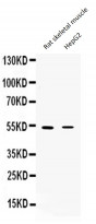
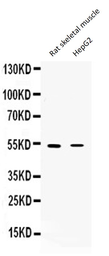
ARG58428 anti-CHRNA5 antibody WB image
Western blot: 50 µg of Rat skeletal muscle and HepG2 whole cell lysate stained with ARG58428 anti-CHRNA5 antibody at 0.5 µg/ml, overnight at 4°C, under reducing conditions.
-
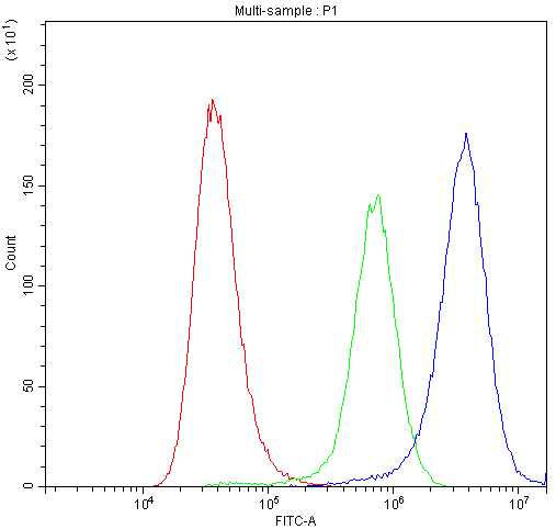
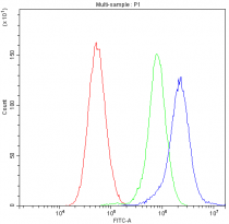
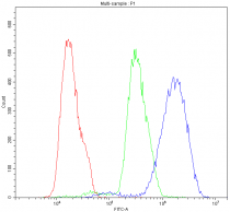
ARG58428 anti-CHRNA5 antibody FACS image
Flow Cytometry: A549 cells were blocked with 10% normal goat serum, and then stained with ARG58428 anti-CHRNA5 antibody (blue) at 1 µg/10^6 cells for 30 min at 20°C, followed by DyLight®488 labelled secondary antibody. Isotype control antibody (green) was rabbit IgG (1 µg/10^6 cells) used under the same conditions. Unlabelled sample (red) was also used as a control.
-
ARG58428 anti-CHRNA5 antibody FACS image
Flow Cytometry: MCF-7 cells were blocked with 10% normal goat serum, and then stained with ARG58428 anti-CHRNA5 antibody (blue) at 1 µg/10^6 cells for 30 min at 20°C, followed by DyLight®488 labelled secondary antibody. Isotype control antibody (green) was rabbit IgG (1 µg/10^6 cells) used under the same conditions. Unlabelled sample (red) was also used as a control.
-
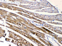
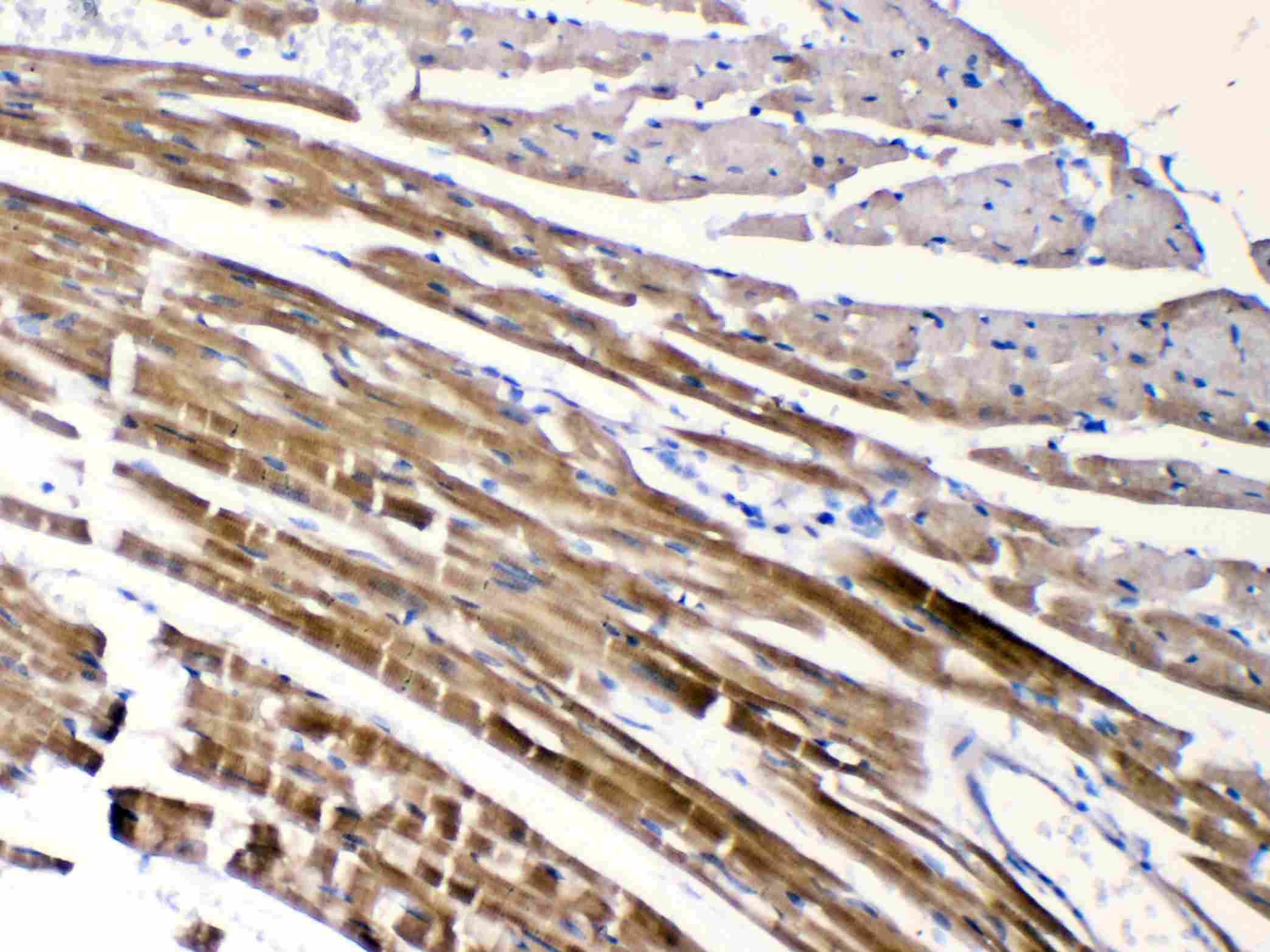
ARG58428 anti-CHRNA5 antibody IHC-P image
Immunohistochemistry: Paraffin-embedded Mouse cardiac muscle tissue. Antigen Retrieval: Heat mediated was performed in Citrate buffer (pH 6.0, epitope retrieval solution) for 20 min. The tissue section was blocked with 10% goat serum. The tissue section was then stained with ARG58428 anti-CHRNA5 antibody at 1 µg/ml dilution, overnight at 4°C.
-
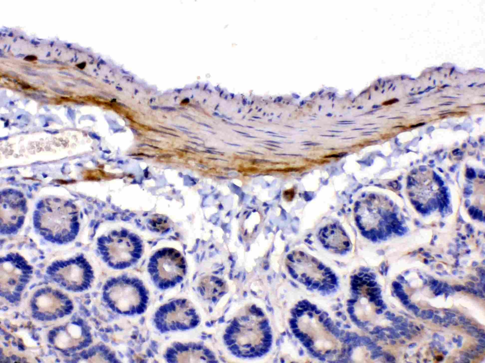
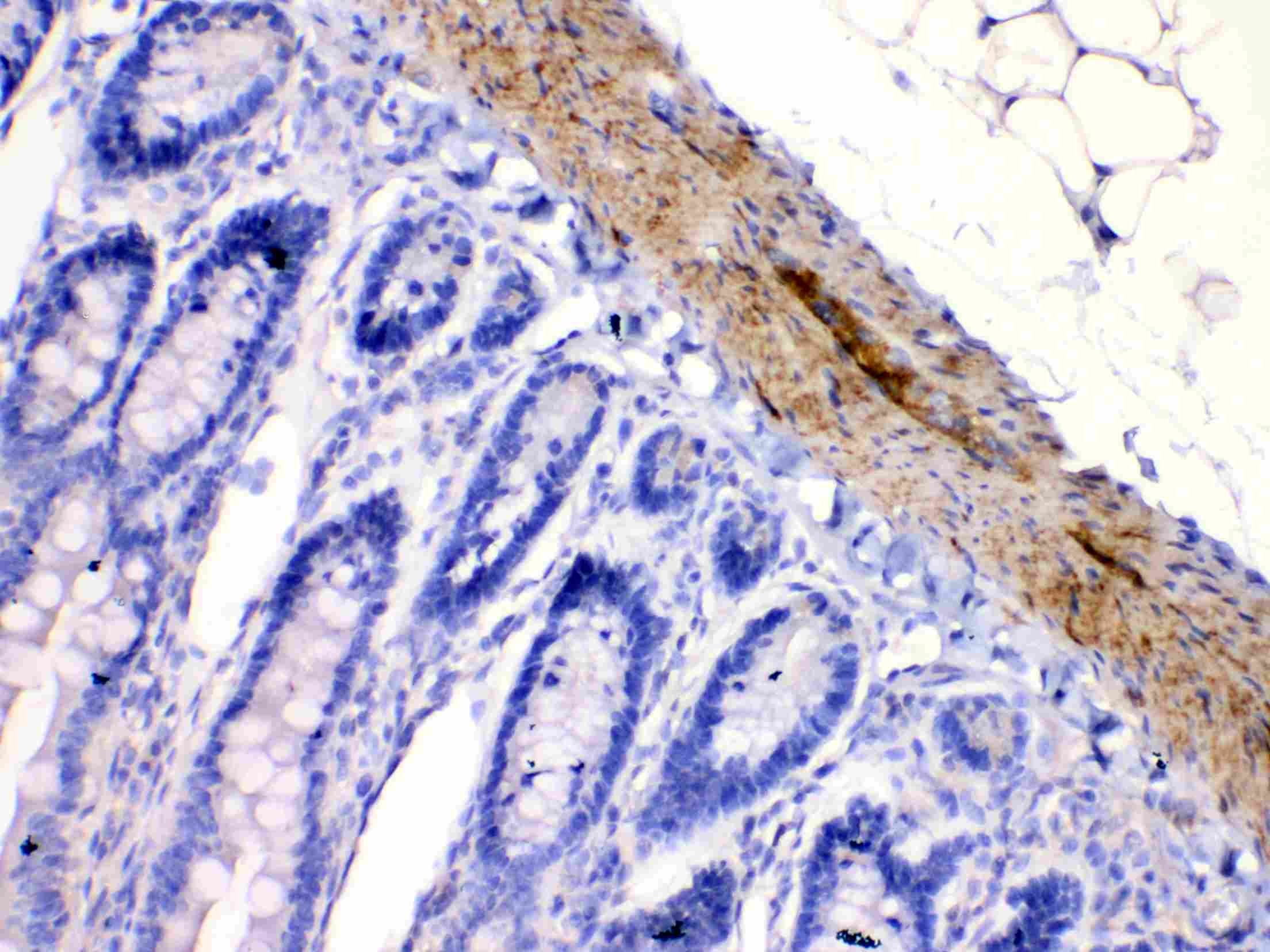
ARG58428 anti-CHRNA5 antibody IHC-P image
Immunohistochemistry: Paraffin-embedded Rat intestine tissue. Antigen Retrieval: Heat mediated was performed in Citrate buffer (pH 6.0, epitope retrieval solution) for 20 min. The tissue section was blocked with 10% goat serum. The tissue section was then stained with ARG58428 anti-CHRNA5 antibody at 1 µg/ml dilution, overnight at 4°C.
-

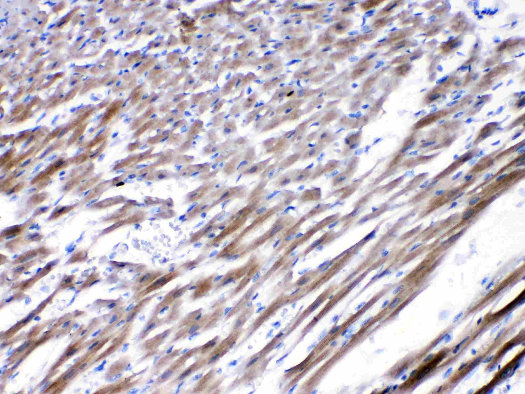
ARG58428 anti-CHRNA5 antibody IHC-P image
Immunohistochemistry: Paraffin-embedded Rat cardiac muscle tissue. Antigen Retrieval: Heat mediated was performed in Citrate buffer (pH 6.0, epitope retrieval solution) for 20 min. The tissue section was blocked with 10% goat serum. The tissue section was then stained with ARG58428 anti-CHRNA5 antibody at 1 µg/ml dilution, overnight at 4°C.
-

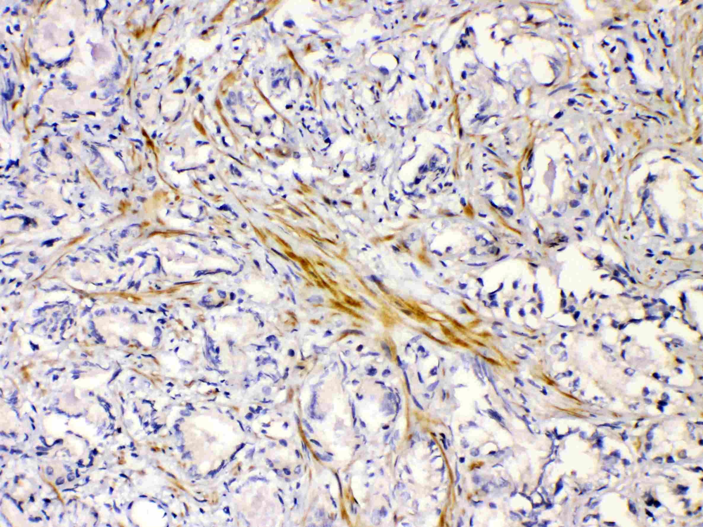
ARG58428 anti-CHRNA5 antibody IHC-P image
Immunohistochemistry: Paraffin-embedded Human prostatic cancer tissue. Antigen Retrieval: Heat mediated was performed in Citrate buffer (pH 6.0, epitope retrieval solution) for 20 min. The tissue section was blocked with 10% goat serum. The tissue section was then stained with ARG58428 anti-CHRNA5 antibody at 1 µg/ml dilution, overnight at 4°C.