ARG58434
anti-CENPA antibody
anti-CENPA antibody for Western blot and Human,Rat
Overview
| Product Description | Rabbit Polyclonal antibody recognizes CENPA |
|---|---|
| Tested Reactivity | Hu, Rat |
| Tested Application | WB |
| Host | Rabbit |
| Clonality | Polyclonal |
| Isotype | IgG |
| Target Name | CENPA |
| Antigen Species | Human |
| Immunogen | Human CENPA recombinant protein (Position: M1-G140). Human CENPA shares 71.6% amino acid (aa) sequence identity with Mouse CENPA. |
| Conjugation | Un-conjugated |
| Alternate Names | CENP-A; Centromere protein A; CenH3; Histone H3-like centromeric protein A; Centromere autoantigen A |
Application Instructions
| Application Suggestion |
|
||||
|---|---|---|---|---|---|
| Application Note | * The dilutions indicate recommended starting dilutions and the optimal dilutions or concentrations should be determined by the scientist. |
Properties
| Form | Liquid |
|---|---|
| Purification | Affinity purification with immunogen. |
| Buffer | 0.9% NaCl, 0.2% Na2HPO4, 0.05% Sodium azide and 5% BSA. |
| Preservative | 0.05% Sodium azide |
| Stabilizer | 5% BSA |
| Concentration | 0.5 mg/ml |
| Storage Instruction | For continuous use, store undiluted antibody at 2-8°C for up to a week. For long-term storage, aliquot and store at -20°C or below. Storage in frost free freezers is not recommended. Avoid repeated freeze/thaw cycles. Suggest spin the vial prior to opening. The antibody solution should be gently mixed before use. |
| Note | For laboratory research only, not for drug, diagnostic or other use. |
Bioinformation
| Database Links |
Swiss-port # P49450 Human Histone H3-like centromeric protein A |
|---|---|
| Gene Symbol | CENPA |
| Gene Full Name | centromere protein A |
| Background | Centromeres are the differentiated chromosomal domains that specify the mitotic behavior of chromosomes. CENPA encodes a centromere protein which contains a histone H3 related histone fold domain that is required for targeting to the centromere. CENPA is proposed to be a component of a modified nucleosome or nucleosome-like structure in which it replaces 1 or both copies of conventional histone H3 in the (H3-H4)2 tetrameric core of the nucleosome particle. Alternative splicing results in multiple transcript variants encoding distinct isoforms. [provided by RefSeq, Jul 2008] |
| Function | Histone H3-like variant which exclusively replaces conventional H3 in the nucleosome core of centromeric chromatin at the inner plate of the kinetochore. Required for recruitment and assembly of kinetochore proteins, mitotic progression and chromosome segregation. May serve as an epigenetic mark that propagates centromere identity through replication and cell division. The CENPA-H4 heterotetramer can bind DNA by itself (in vitro). [UniProt] |
| Cellular Localization | Nucleus. Chromosome, centromere, kinetochore. Localizes exclusively in the kinetochore domain of centromeres. Occupies a compact domain at the inner kinetochore plate stretching across 2 thirds of the length of the constriction but encompassing only one third of the constriction width and height. [UniProt] |
| Calculated MW | 16 kDa |
| PTM | Ubiquitinated (Probable). Interaction with herpes virus HSV-1 ICP0 protein, leads to its degradation by the proteasome pathway. Trimethylated by NTMT1 at the N-terminal glycine after cleavage of Met-1. Methylation is low before incorporation into nucleosomes and increases with cell cycle progression, with the highest levels in mitotic nucleosomes. Phosphorylated by CDK1 at Ser-68 during early mitosis; this abolishes association with chromatin and centromeres, prevents interaction with HJURP and thereby prevents premature assembly of CENPA into centromeres (PubMed:25556658). Dephosphorylated at Ser-68 by PPP1CA during late mitosis (PubMed:25556658). Phosphorylation of Ser-7 by AURKA and AURKB during prophase is required for localization of AURKA and AURKB at inner centromere and is essential for normal cytokinesis (PubMed:11756469, PubMed:14667408, PubMed:18239465). Initial phosphorylation during prophase is mediated by AURKA and is maintained by AURKB. Poly-ADP-ribosylated by PARP1. [UniProt] |
Images (1) Click the Picture to Zoom In
-
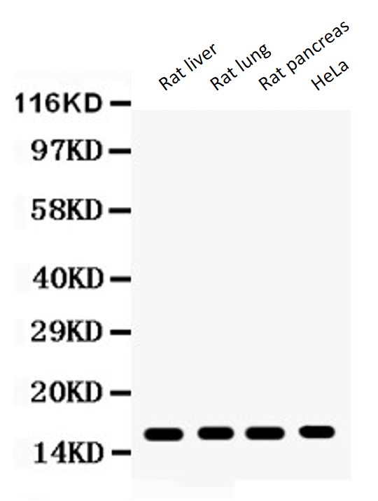
ARG58434 anti-CENPA antibody WB image
Western blot: 50 µg of Rat liver, 50 µg of Rat lung, 50 µg of Rat pancreas and 40 µg of HeLa whole cell lysate stained with ARG58434 anti-CENPA antibody at 0.5 µg/ml dilution.