ARG55212
anti-CDK5 antibody
anti-CDK5 antibody for Flow cytometry,ICC/IF,Western blot and Human,Mouse,Rat
Cancer antibody; Cell Biology and Cellular Response antibody; Metabolism antibody; Neuroscience antibody
Overview
| Product Description | Rabbit Polyclonal antibody recognizes CDK5 |
|---|---|
| Tested Reactivity | Hu, Ms, Rat |
| Tested Application | FACS, ICC/IF, WB |
| Host | Rabbit |
| Clonality | Polyclonal |
| Isotype | IgG |
| Target Name | CDK5 |
| Antigen Species | Human |
| Immunogen | KLH-conjugated synthetic peptide corresponding to aa. 254-289 (C-terminus) of Human CDK5. |
| Conjugation | Un-conjugated |
| Alternate Names | Cell division protein kinase 5; TPKII catalytic subunit; LIS7; PSSALRE; Serine/threonine-protein kinase PSSALRE; Cyclin-dependent-like kinase 5; EC 2.7.11.1; Tau protein kinase II catalytic subunit |
Application Instructions
| Application Suggestion |
|
||||||||
|---|---|---|---|---|---|---|---|---|---|
| Application Note | * The dilutions indicate recommended starting dilutions and the optimal dilutions or concentrations should be determined by the scientist. |
Properties
| Form | Liquid |
|---|---|
| Purification | Purification with Protein A and immunogen peptide. |
| Buffer | PBS and 0.09% (W/V) Sodium azide |
| Preservative | 0.09% (W/V) Sodium azide |
| Storage Instruction | For continuous use, store undiluted antibody at 2-8°C for up to a week. For long-term storage, aliquot and store at -20°C or below. Storage in frost free freezers is not recommended. Avoid repeated freeze/thaw cycles. Suggest spin the vial prior to opening. The antibody solution should be gently mixed before use. |
| Note | For laboratory research only, not for drug, diagnostic or other use. |
Bioinformation
| Database Links | |
|---|---|
| Gene Symbol | CDK5 |
| Gene Full Name | cyclin-dependent kinase 5 |
| Background | This gene encodes a proline-directed serine/threonine kinase that is a member of the cyclin-dependent kinase family of proteins. Unlike other members of the family, the protein encoded by this gene does not directly control cell cycle regulation. Instead the protein, which is predominantly expressed at high levels in mammalian postmitotic central nervous system neurons, functions in diverse processes such as synaptic plasticity and neuronal migration through phosphorylation of proteins required for cytoskeletal organization, endocytosis and exocytosis, and apoptosis. In humans, an allelic variant of the gene that results in undetectable levels of the protein has been associated with lethal autosomal recessive lissencephaly-7. Alternative splicing results in multiple transcript variants. [provided by RefSeq, May 2015] |
| Function | Proline-directed serine/threonine-protein kinase essential for neuronal cell cycle arrest and differentiation and may be involved in apoptotic cell death in neuronal diseases by triggering abortive cell cycle re-entry. Interacts with D1 and D3-type G1 cyclins. Phosphorylates SRC, NOS3, VIM/vimentin, p35/CDK5R1, MEF2A, SIPA1L1, SH3GLB1, PXN, PAK1, MCAM/MUC18, SEPT5, SYN1, DNM1, AMPH, SYNJ1, CDK16, RAC1, RHOA, CDC42, TONEBP/NFAT5, MAPT/TAU, MAP1B, histone H1, p53/TP53, HDAC1, APEX1, PTK2/FAK1, huntingtin/HTT, ATM, MAP2, NEFH and NEFM. Regulates several neuronal development and physiological processes including neuronal survival, migration and differentiation, axonal and neurite growth, synaptogenesis, oligodendrocyte differentiation, synaptic plasticity and neurotransmission, by phosphorylating key proteins. Activated by interaction with CDK5R1 (p35) and CDK5R2 (p39), especially in post-mitotic neurons, and promotes CDK5R1 (p35) expression in an autostimulation loop. Phosphorylates many downstream substrates such as Rho and Ras family small GTPases (e.g. PAK1, RAC1, RHOA, CDC42) or microtubule-binding proteins (e.g. MAPT/TAU, MAP2, MAP1B), and modulates actin dynamics to regulate neurite growth and/or spine morphogenesis. Phosphorylates also exocytosis associated proteins such as MCAM/MUC18, SEPT5, SYN1, and CDK16/PCTAIRE1 as well as endocytosis associated proteins such as DNM1, AMPH and SYNJ1 at synaptic terminals. [UniProt] |
| Cellular Localization | Cell membrane, Cell projection, Cytoplasm, Membrane, Nucleus, Synapse |
| Research Area | Cancer antibody; Cell Biology and Cellular Response antibody; Metabolism antibody; Neuroscience antibody |
| Calculated MW | 33 kDa |
| PTM | Phosphorylation on Tyr-15 by ABL1 and FYN, and on Ser-159 by casein kinase 1 promotes kinase activity. By contrast, phosphorylation at Thr-14 inhibits activity. Phosphorylation at Ser-159 is essential for maximal catalytic activity. |
Images (3) Click the Picture to Zoom In
-

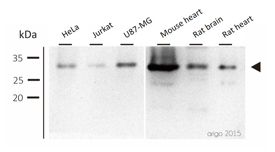
ARG55212 anti-CDK5 antibody WB image
Western blot: 30 μg of HeLa, Jurkat, U87-MG, Mouse heart, Rat brain and Rat heart lysates stained with ARG55212 anti-CDK5 antibody at 1:500 dilution.
-
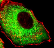
ARG55212 anti-CDK5 antibody ICC/IF image
Immunofluorescence: A549 cells stained with ARG55212 anti-CDK5 antibody (green) at 1:25 dilution. Cytoplasmic actin was counterstained with Alexa Fluor® 555 conjugated with Phalloidin (red).
-
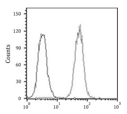
ARG55212 anti-CDK5 antibody FACS image
Flow Cytometry: K562 cells stained with ARG55212 anti-CDK5 antibody (right histogram) at 1:25 dilution or without primary antibodies (left histogram), followed by incubation with Alexa Fluor® 488 labelled secondary antibody.