ARG66317
anti-CDK4 antibody [SQab1865]
anti-CDK4 antibody [SQab1865] for Western blot and Human,Mouse,African green monkey,Bovine,Dog,Pig
Overview
| Product Description | Recombinant Rabbit Monoclonal antibody [SQab1865] recognizes CDK4 |
|---|---|
| Tested Reactivity | Hu, Ms, AGMK, Bov, Dog, Pig |
| Tested Application | WB |
| Host | Rabbit |
| Clonality | Monoclonal |
| Clone | SQab1865 |
| Isotype | IgG |
| Target Name | CDK4 |
| Antigen Species | Human |
| Immunogen | Synthetic peptide around the N-terminus of CDK4. |
| Conjugation | Un-conjugated |
| Alternate Names | Cyclin-dependent kinase 4; PSK-J3; CMM3; EC 2.7.11.22; Cell division protein kinase 4 |
Application Instructions
| Application Suggestion |
|
||||
|---|---|---|---|---|---|
| Application Note | * The dilutions indicate recommended starting dilutions and the optimal dilutions or concentrations should be determined by the scientist. |
Properties
| Form | Liquid |
|---|---|
| Purification | Purification with Protein A. |
| Buffer | PBS, 0.01% Sodium azide, 40% Glycerol and 0.05% BSA. |
| Preservative | 0.01% Sodium azide |
| Stabilizer | 40% Glycerol and 0.05% BSA |
| Storage Instruction | For continuous use, store undiluted antibody at 2-8°C for up to a week. For long-term storage, aliquot and store at -20°C. Storage in frost free freezers is not recommended. Avoid repeated freeze/thaw cycles. Suggest spin the vial prior to opening. The antibody solution should be gently mixed before use. |
| Note | For laboratory research only, not for drug, diagnostic or other use. |
Bioinformation
| Database Links | |
|---|---|
| Gene Symbol | CDK4 |
| Gene Full Name | cyclin-dependent kinase 4 |
| Background | The protein encoded by this gene is a member of the Ser/Thr protein kinase family. This protein is highly similar to the gene products of S. cerevisiae cdc28 and S. pombe cdc2. It is a catalytic subunit of the protein kinase complex that is important for cell cycle G1 phase progression. The activity of this kinase is restricted to the G1-S phase, which is controlled by the regulatory subunits D-type cyclins and CDK inhibitor p16(INK4a). This kinase was shown to be responsible for the phosphorylation of retinoblastoma gene product (Rb). Mutations in this gene as well as in its related proteins including D-type cyclins, p16(INK4a) and Rb were all found to be associated with tumorigenesis of a variety of cancers. Multiple polyadenylation sites of this gene have been reported. [provided by RefSeq, Jul 2008] |
| Function | Ser/Thr-kinase component of cyclin D-CDK4 (DC) complexes that phosphorylate and inhibit members of the retinoblastoma (RB) protein family including RB1 and regulate the cell-cycle during G(1)/S transition. Phosphorylation of RB1 allows dissociation of the transcription factor E2F from the RB/E2F complexes and the subsequent transcription of E2F target genes which are responsible for the progression through the G(1) phase. Hypophosphorylates RB1 in early G(1) phase. Cyclin D-CDK4 complexes are major integrators of various mitogenenic and antimitogenic signals. Also phosphorylates SMAD3 in a cell-cycle-dependent manner and represses its transcriptional activity. Component of the ternary complex, cyclin D/CDK4/CDKN1B, required for nuclear translocation and activity of the cyclin D-CDK4 complex. [UniProt] |
| Calculated MW | 34 kDa |
| PTM | Phosphorylation at Thr-172 is required for enzymatic activity. Phosphorylated, in vitro, at this site by CCNH-CDK7, but, in vivo, appears to be phosphorylated by a proline-directed kinase. In the cyclin D-CDK4-CDKN1B complex, this phosphorylation and consequent CDK4 enzyme activity, is dependent on the tyrosine phosphorylation state of CDKN1B. Thus, in proliferating cells, CDK4 within the complex is phosphorylated on Thr-172 in the T-loop. In resting cells, phosphorylation on Thr-172 is prevented by the non-tyrosine-phosphorylated form of CDKN1B. [UniProt] |
Images (4) Click the Picture to Zoom In
-
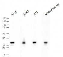
ARG66317 anti-CDK4 antibody [SQab1865] WB image
Western blot: 10 µg of HeLa, K562, 3T3 and Mouse kidney lysates stained with ARG66317 anti-CDK4 antibody [SQab1865] at 1:1000 dilution.
-
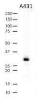
ARG66317 anti-CDK4 antibody [SQab1865] WB image
Western blot: 10 µg of A431 cell lysates stained with ARG66317 anti-CDK4 antibody [SQab1865] at 1:1000 dilution.
-
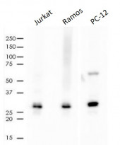
ARG66317 anti-CDK4 antibody [SQab1865] WB image
Western blot: 10 µg of Jurkat, Ramos and PC-12 cell lysates stained with ARG66317 anti-CDK4 antibody [SQab1865] at 1:500 dilution.
-
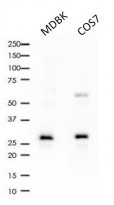
ARG66317 anti-CDK4 antibody [SQab1865] WB image
Western blot: 10 µg of MDBK and COS7 cell lysates stained with ARG66317 anti-CDK4 antibody [SQab1865] at 1:1000 dilution.


