ARG63499
anti-CDCP1 antibody
anti-CDCP1 antibody for IHC-Formalin-fixed paraffin-embedded sections,Western blot and Human
Cancer antibody; Controls and Markers antibody; Developmental Biology antibody
Overview
| Product Description | Goat Polyclonal antibody recognizes CDCP1 |
|---|---|
| Tested Reactivity | Hu |
| Tested Application | IHC-P, WB |
| Specificity | This antibody is expected to recognize isoform 1 (NP_073753.3) only. |
| Host | Goat |
| Clonality | Polyclonal |
| Isotype | IgG |
| Target Name | CDCP1 |
| Antigen Species | Human |
| Immunogen | PLLNTQEPMEPAE |
| Conjugation | Un-conjugated |
| Alternate Names | Membrane glycoprotein gp140; CD antigen CD318; SIMA135; Subtractive immunization M plus HEp3-associated 135 kDa protein; CUB domain-containing protein 1; TRASK; Transmembrane and associated with src kinases; CD318 |
Application Instructions
| Application Suggestion |
|
||||||
|---|---|---|---|---|---|---|---|
| Application Note | WB: Recommend incubate at RT for 1h. IHC-P: Antigen Retrieval: Steam tissue section in Citrate buffer (pH 6.0). * The dilutions indicate recommended starting dilutions and the optimal dilutions or concentrations should be determined by the scientist. |
Properties
| Form | Liquid |
|---|---|
| Purification | Purified from goat serum by antigen affinity chromatography. |
| Buffer | Tris saline (pH 7.3), 0.02% Sodium azide and 0.5% BSA. |
| Preservative | 0.02% Sodium azide |
| Stabilizer | 0.5% BSA |
| Concentration | 0.5 mg/ml |
| Storage Instruction | For continuous use, store undiluted antibody at 2-8°C for up to a week. For long-term storage, aliquot and store at -20°C or below. Storage in frost free freezers is not recommended. Avoid repeated freeze/thaw cycles. Suggest spin the vial prior to opening. The antibody solution should be gently mixed before use. |
| Note | For laboratory research only, not for drug, diagnostic or other use. |
Bioinformation
| Database Links | |
|---|---|
| Background | The protein encoded by this gene is a transmembrane protein containing three extracellular CUB domains. This protein is found to be overexpressed in colon and lung cancers. Its expression level is correlated with the metastatic ability of carcinoma cells. This protein is located on the cell surface. It has been shown to be tyrosine phosphorylated in a cancer cell line. Alternatively spliced transcript variants encoding distinct isoforms have been reported. [provided by RefSeq, Jul 2008] |
| Research Area | Cancer antibody; Controls and Markers antibody; Developmental Biology antibody |
| Calculated MW | 93 kDa |
| PTM | Phosphorylated on tyrosine by kinases of the SRC family such as SRC and YES as well as by the protein kinase C gamma/PRKCG. Dephosphorylated by phosphotyrosine phosphatases. Also phosphorylated by suramin, a heparin analog. Tyrosine phosphorylated in response to dissociation of integrin alpha-6 beta-4 from laminin-5. N-glycosylated. A soluble form may also be produced by proteolytic cleavage at the cell surface (shedding). Another peptide of 80 kDa (p80) is present in cultured keratinocytes probably due to tryptic cleavage at an unidentified site on its N-terminal side. Converted to p80 by plasmin, a trypsin-like protease. |
Images (3) Click the Picture to Zoom In
-
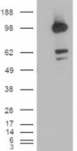
ARG63499 anti-CDCP1 antibody WB image
Western Blot: Human Colon lysate (35 µg protein in RIPA buffer) stained with ARG63499 anti-CDCP1 antibody at 1 µg/ml dilution.
-
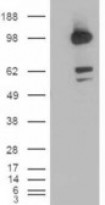
ARG63499 anti-CDCP1 antibody WB image
Western Blot: 1). Mock transfection; 2) CDCP1 (RC220633) expressing plasmid transfected HEK293 cell lysate standed with ARG63499 anti-CDCP1 antibody
-
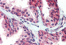
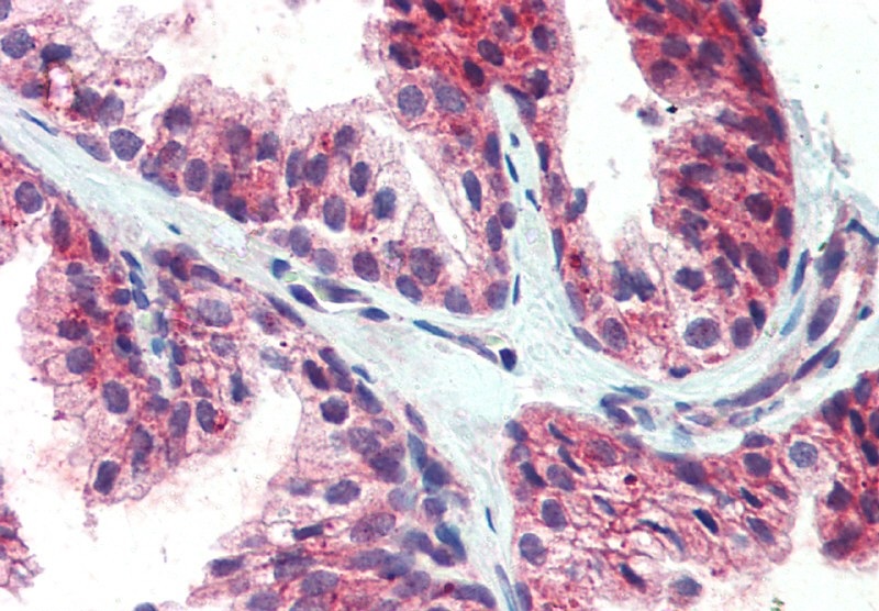
ARG63499 anti-CDCP1 antibody IHC-P image
Immunohistochemistry: Paraffin-embedded Human prostate tissue. Antigen Retrieval: Steam tissue section in Citrate buffer (pH 6.0). The tissue section was stained with ARG63499 anti-CDCP1 antibody at 5 µg/ml dilution followed by AP-staining.