ARG65389
anti-CD99 antibody [3B2/TA8]
anti-CD99 antibody [3B2/TA8] for CyTOF®-candidate,Flow cytometry and Human
Cancer antibody; Immune System antibody; Signaling Transduction antibody
Overview
| Product Description | Mouse Monoclonal antibody [3B2/TA8] recognizes CD99 |
|---|---|
| Tested Reactivity | Hu |
| Tested Application | CyTOF®-candidate, FACS |
| Specificity | The clone 3B2/TA8 recognizes CD99, an approximately 32 kDa sialoglycoprotein expressed on many cell types, with particularly strong expression on Ewing´s sarcoma and peripheral primitive neuroectodermal tumors. Within the hematopoietic system, CD99 is expressed on virtually all cell types except granulocytes. HLDA VI.; WS Code T 6T-097, BP 534 |
| Host | Mouse |
| Clonality | Monoclonal |
| Clone | 3B2/TA8 |
| Isotype | IgG2a |
| Target Name | CD99 |
| Antigen Species | Human |
| Immunogen | Human thymocytes |
| Conjugation | Un-conjugated |
| Alternate Names | 12E7; CD99 antigen; MIC2X; MIC2Y; CD antigen CD99; MSK5X; Protein MIC2; MIC2; T-cell surface glycoprotein E2; HBA71; E2 antigen |
Application Instructions
| Application Suggestion |
|
||||||
|---|---|---|---|---|---|---|---|
| Application Note | * The dilutions indicate recommended starting dilutions and the optimal dilutions or concentrations should be determined by the scientist. |
Properties
| Form | Liquid |
|---|---|
| Purification | Purified from cell culture supernatant by protein-A affinity chromatography. |
| Purity | > 95% (by SDS-PAGE) |
| Buffer | PBS (pH 7.4) and 15 mM Sodium azide |
| Preservative | 15 mM Sodium azide |
| Concentration | 1 mg/ml |
| Storage Instruction | For continuous use, store undiluted antibody at 2-8°C for up to a week. For long-term storage, aliquot and store at -20°C or below. Storage in frost free freezers is not recommended. Avoid repeated freeze/thaw cycles. Suggest spin the vial prior to opening. The antibody solution should be gently mixed before use. |
| Note | For laboratory research only, not for drug, diagnostic or other use. |
Bioinformation
| Database Links | |
|---|---|
| Gene Symbol | CD99 |
| Gene Full Name | CD99 molecule |
| Background | CD99 is a cell surface glycoprotein involved in leukocyte migration, T-cell adhesion, ganglioside GM1 and transmembrane protein transport, and T-cell death by a caspase-independent pathway. In addition, the encoded protein may have the ability to rearrange the actin cytoskeleton and may also act as an oncosuppressor in osteosarcoma. This gene is found in the pseudoautosomal region of chromosomes X and Y and escapes X-chromosome inactivation. There is a related pseudogene located immediately adjacent to this locus. [provided by RefSeq, Mar 2016] |
| Function | CD99 involved in T-cell adhesion processes and in spontaneous rosette formation with erythrocytes. Plays a role in a late step of leukocyte extravasation helping leukocytes to overcome the endothelial basement membrane. Acts at the same site as, but independently of, PECAM1. Involved in T-cell adhesion processes. [UniProt] |
| Research Area | Cancer antibody; Immune System antibody; Signaling Transduction antibody |
| Calculated MW | 19 kDa |
| PTM | Extensively O-glycosylated. |
Images (3) Click the Picture to Zoom In
-
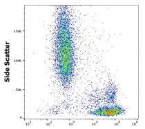
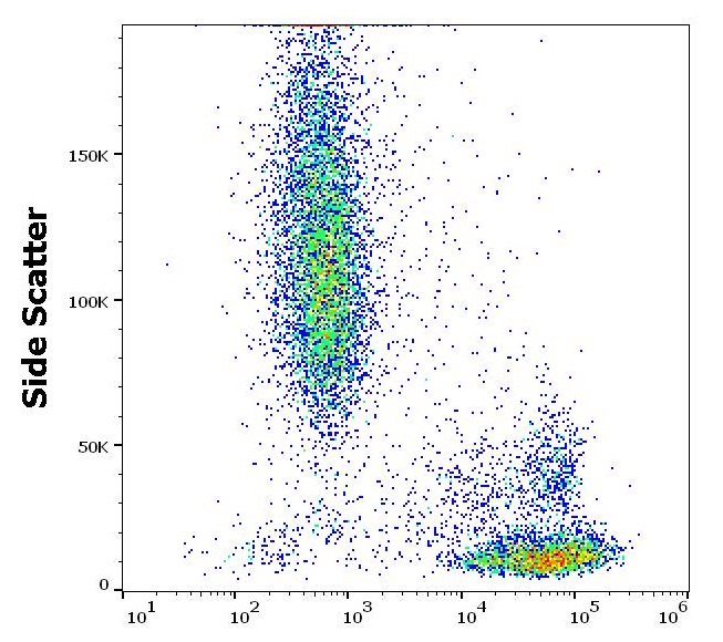
ARG65389 anti-CD99 antibody [3B2/TA8] FACS image
Flow Cytometry: Human peripheral blood stained with ARG65389 anti-CD99 antibody [3B2/TA8] at 2 µg/ml dilution, followed by APC-conjugated Goat anti-Mouse antibody.
-
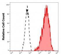
ARG65389 anti-CD99 antibody [3B2/TA8] FACS image
Flow Cytometry: Separation of human lymphocytes (red-filled) from neutrophil granulocytes (black-dashed). Human peripheral whole blood stained with ARG65389 anti-CD99 antibody [3B2/TA8] at 2 µg/ml dilution, followed by APC-conjugated Goat anti-Mouse antibody.
-

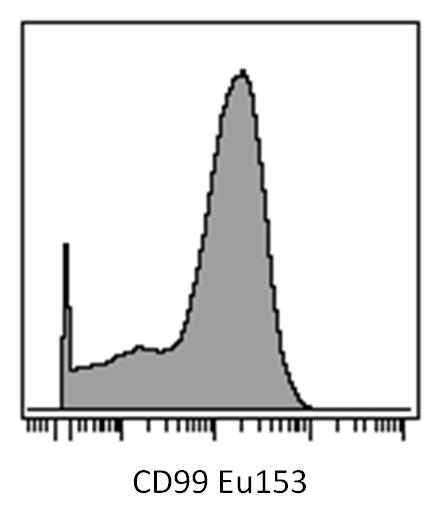
ARG65389 anti-CD99 antibody [3B2/TA8] CyTOF image
CyTOF: Human peripheral blood cells (after Ficoll-Paque separation) stained with ARG65389 anti-CD99 antibody [3B2/TA8] (Eu153). Singlet cells were gated for data analysis.

Clone References
Lipid rafts and pseudotyping.


CD99 engagement on human peripheral blood T cells results in TCR/CD3-dependent cellular activation and allows for Th1-restricted cytokine production.


