ARG62951
anti-CD95 / Fas antibody [LT95]
anti-CD95 / Fas antibody [LT95] for Flow cytometry,IHC-Formalin-fixed paraffin-embedded sections and Human
Cell Biology and Cellular Response antibody; Cell Death antibody; Immune System antibody
Overview
| Product Description | Mouse Monoclonal antibody [LT95] recognizes CD95 / Fas |
|---|---|
| Tested Reactivity | Hu |
| Tested Application | FACS, IHC-P |
| Specificity | The clone LT95 reacts with CD95 (Fas/APO-1), a 46 kDa single chain type I glycoprotein of the tumour necrosis factor/nerve growth factor (TNF/NGF) receptor superfamily, expressed on a variety of normal and neoplastic cells. It seems that the antibody LT95 does not induce Fas mediated apoptosis, although it cross-blocks anti-Fas DX2 antibody that recognizes a functional epitope of Fas molecule. |
| Host | Mouse |
| Clonality | Monoclonal |
| Clone | LT95 |
| Isotype | IgG1 |
| Target Name | CD95 / Fas |
| Antigen Species | Human |
| Immunogen | HUT-78 human T cell lymphoma cell line |
| Conjugation | Un-conjugated |
| Alternate Names | CD95; Apoptosis-mediating surface antigen FAS; FAS1; Tumor necrosis factor receptor superfamily member 6; ALPS1A; APT1; FASTM; CD antigen CD95; APO-1; TNFRSF6; FASLG receptor; Apo-1 antigen |
Application Instructions
| Application Suggestion |
|
||||||
|---|---|---|---|---|---|---|---|
| Application Note | * The dilutions indicate recommended starting dilutions and the optimal dilutions or concentrations should be determined by the scientist. | ||||||
| Positive Control | IHC-P: Tonsil |
Properties
| Form | Liquid |
|---|---|
| Purification | Purified from cell culture supernatant by protein-A affinity chromatography. |
| Purity | > 95% (by SDS-PAGE) |
| Buffer | PBS (pH 7.4) and 15 mM Sodium azide |
| Preservative | 15 mM Sodium azide |
| Concentration | 1 mg/ml |
| Storage Instruction | For continuous use, store undiluted antibody at 2-8°C for up to a week. For long-term storage, aliquot and store at -20°C or below. Storage in frost free freezers is not recommended. Avoid repeated freeze/thaw cycles. Suggest spin the vial prior to opening. The antibody solution should be gently mixed before use. |
| Note | For laboratory research only, not for drug, diagnostic or other use. |
Bioinformation
| Database Links |
Swiss-port # P25445 Human Tumor necrosis factor receptor superfamily member 6 |
|---|---|
| Gene Symbol | FAS |
| Gene Full Name | Fas cell surface death receptor |
| Background | CD95 (Fas, APO-1), a 46 kDa transmembrane glycoprotein, is a cell death receptor of the TNFR superfamily. Stimulation of CD95 results in aggregation of its intracellular death domains, formation of the death-inducing signaling complex (DISC) and activation of caspases. In type I cells caspase 3 is activated by high amounts of caspase 8 generated at the DISC, in type II cells low concentration of caspase 8 activates pathway leading to the release of cytochrome c from mitochondria and activation of caspase 3 by cytochom c. Besides its roles in induction of apoptosis, Fas also triggers pro-inflammatory cytokine responses. |
| Function | Receptor for TNFSF6/FASLG. The adapter molecule FADD recruits caspase-8 to the activated receptor. The resulting death-inducing signaling complex (DISC) performs caspase-8 proteolytic activation which initiates the subsequent cascade of caspases (aspartate-specific cysteine proteases) mediating apoptosis. FAS-mediated apoptosis may have a role in the induction of peripheral tolerance, in the antigen-stimulated suicide of mature T-cells, or both. The secreted isoforms 2 to 6 block apoptosis (in vitro). [UniProt] |
| Research Area | Cell Biology and Cellular Response antibody; Cell Death antibody; Immune System antibody |
| Calculated MW | 38 kDa |
| PTM | N- and O-glycosylated. O-glycosylated with core 1 or possibly core 8 glycans. |
Images (1) Click the Picture to Zoom In
-
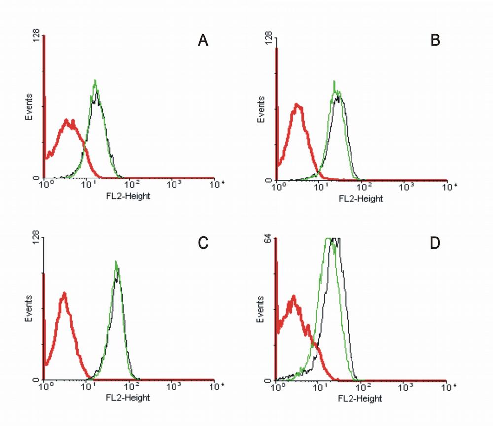
ARG62951 anti-CD95 / Fas antibody [LT95] FACS image
Flow Cytometry: A) Jurkat cells, B) RAMOS cells, C) CEM human leukemia cells, and D) MOLT-4 cells stained with ARG62951 anti-CD95 / Fas antibody [LT95], followed by incubation with PE-labelled secondary antibody.
Clone References
Transformation by oncogenic RAS sensitizes human colon cells to TRAIL-induced apoptosis by up-regulating death receptor 4 and death receptor 5 through a MEK-dependent pathway.


