ARG42258
anti-CD86 antibody [BU63] (low endotoxin)
anti-CD86 antibody [BU63] (low endotoxin) for Flow cytometry,Functional study,IHC-Frozen sections,Immunoprecipitation,Western blot and Human
Overview
| Product Description | Azide free and low endotoxin Mouse Monoclonal antibody [BU63] recognizes CD86 |
|---|---|
| Tested Reactivity | Hu |
| Tested Application | FACS, FuncSt, IHC-Fr, IP, WB |
| Specificity | The mouse monoclonal antibody BU63 reacts with an extracellular epitope of CD86 (B7-2), a 70 kDa type I transmembrane glycoprotein of immunoglobulin supergene family, expressed on professional antigen-presenting cells, such as dendritic cells, macrophages or activated B lymphocytes. |
| Host | Mouse |
| Clonality | Monoclonal |
| Clone | BU63 |
| Isotype | IgG1 |
| Target Name | CD86 |
| Antigen Species | Human |
| Immunogen | B-lymphoblastoid cell line ARH-77. |
| Conjugation | Un-conjugated |
| Alternate Names | B70; B7.2; LAB72; CD antigen CD86; B7-2; FUN-1; CD28LG2; T-lymphocyte activation antigen CD86; CTLA-4 counter-receptor B7.2; Activation B7-2 antigen; BU63 |
Application Instructions
| Application Suggestion |
|
||||||||||||
|---|---|---|---|---|---|---|---|---|---|---|---|---|---|
| Application Note | * The dilutions indicate recommended starting dilutions and the optimal dilutions or concentrations should be determined by the scientist. |
Properties
| Form | Liquid |
|---|---|
| Purification | Purification with Protein A. |
| Purification Note | 0.2 µm filter sterilized. Endotoxin level is <0.01 EU/µg of the protein. |
| Buffer | PBS |
| Concentration | 1 mg/ml |
| Storage Instruction | For continuous use, store undiluted antibody at 2-8°C for up to a week. For long-term storage, aliquot and store at -20°C or below. Storage in frost free freezers is not recommended. Avoid repeated freeze/thaw cycles. Suggest spin the vial prior to opening. The antibody solution should be gently mixed before use. |
| Note | For laboratory research only, not for drug, diagnostic or other use. |
Bioinformation
| Database Links |
Swiss-port # P42081 Human T-lymphocyte activation antigen CD86 |
|---|---|
| Gene Symbol | CD86 |
| Gene Full Name | CD86 molecule |
| Background | This gene encodes a type I membrane protein that is a member of the immunoglobulin superfamily. This protein is expressed by antigen-presenting cells, and it is the ligand for two proteins at the cell surface of T cells, CD28 antigen and cytotoxic T-lymphocyte-associated protein 4. Binding of this protein with CD28 antigen is a costimulatory signal for activation of the T-cell. Binding of this protein with cytotoxic T-lymphocyte-associated protein 4 negatively regulates T-cell activation and diminishes the immune response. Alternative splicing results in several transcript variants encoding different isoforms. [provided by RefSeq, May 2011] |
| Function | Receptor involved in the costimulatory signal essential for T-lymphocyte proliferation and interleukin-2 production, by binding CD28 or CTLA-4. May play a critical role in the early events of T-cell activation and costimulation of naive T-cells, such as deciding between immunity and anergy that is made by T-cells within 24 hours after activation. Isoform 2 interferes with the formation of CD86 clusters, and thus acts as a negative regulator of T-cell activation. (Microbial infection) Acts as a receptor for adenovirus subgroup B. [UniProt] |
| Cellular Localization | Cell membrane; Single-pass type I membrane protein. [UniProt] |
| Calculated MW | 38 kDa |
| PTM | Polyubiquitinated; which is promoted by MARCH8 and results in endocytosis and lysosomal degradation. [UniProt] |
Images (2) Click the Picture to Zoom In
-

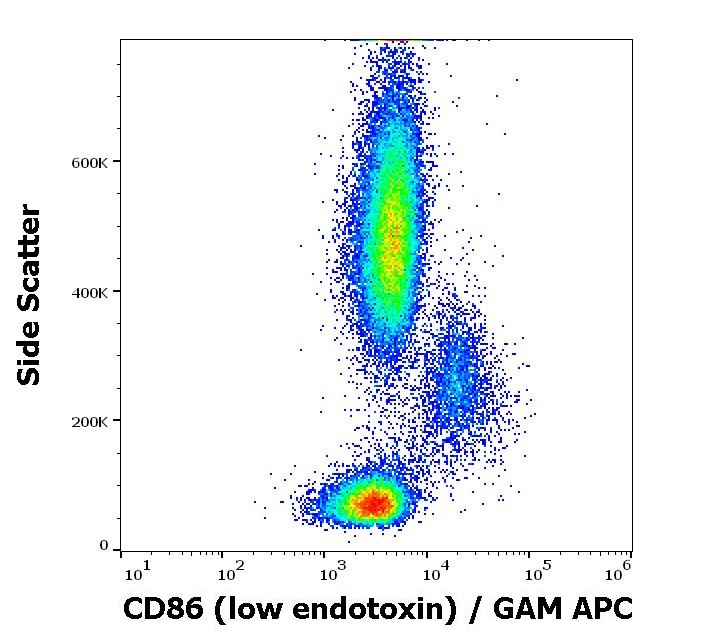
ARG42258 anti-CD86 antibody [BU63] (low endotoxin) FACS image
Flow Cytometry: Human peripheral blood stained with ARG42258 anti-CD86 antibody [BU63] (low endotoxin) at 3 µg/ml dilution, followed by APC-conjugated Goat anti-Mouse antibody.
-

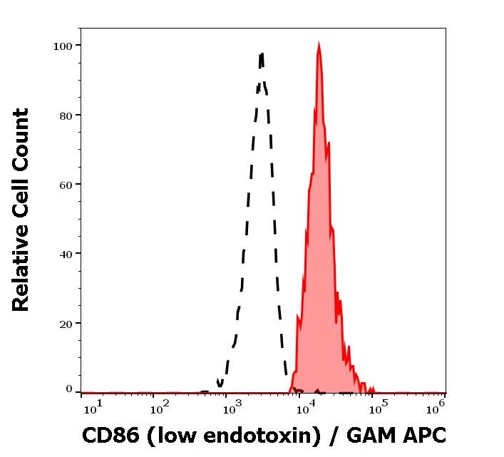
ARG42258 anti-CD86 antibody [BU63] (low endotoxin) FACS image
Flow Cytometry: Separation of Human monocytes (red-filled) from lymphocytes (black-dashed). Human peripheral whole blood stained with ARG42258 anti-CD86 antibody [BU63] (low endotoxin) at 3 µg/ml dilution, followed by APC-conjugated Goat anti-Mouse antibody.