ARG40691
anti-CD81 antibody
anti-CD81 antibody for Flow cytometry,ICC/IF,IHC-Formalin-fixed paraffin-embedded sections,Western blot and Mouse
Overview
| Product Description | Rabbit Polyclonal antibody recognizes CD81 |
|---|---|
| Tested Reactivity | Ms |
| Tested Application | FACS, ICC/IF, IHC-P, WB |
| Host | Rabbit |
| Clonality | Polyclonal |
| Isotype | IgG |
| Target Name | CD81 |
| Antigen Species | Mouse |
| Immunogen | Recombinant protein corresponding to K116-K201 of Mouse CD81. |
| Conjugation | Un-conjugated |
| Alternate Names | CD antigen CD81; TAPA1; Tspan-28; S5.7; CD81 antigen; Target of the antiproliferative antibody 1; Tetraspanin-28; 26 kDa cell surface protein TAPA-1; CVID6; TSPAN28 |
Application Instructions
| Application Suggestion |
|
||||||||||
|---|---|---|---|---|---|---|---|---|---|---|---|
| Application Note | IHC-P: Antigen Retrieval: Heat mediation was performed in EDTA buffer (pH 8.0). * The dilutions indicate recommended starting dilutions and the optimal dilutions or concentrations should be determined by the scientist. |
Properties
| Form | Liquid |
|---|---|
| Buffer | 0.2% Na2HPO4, 0.9% NaCl, 0.05% Sodium azide and 4% Trehalose. |
| Preservative | 0.05% Sodium azide |
| Stabilizer | 4% Trehalose |
| Concentration | 0.5 mg/ml |
| Storage Instruction | For continuous use, store undiluted antibody at 2-8°C for up to a week. For long-term storage, aliquot and store at -20°C or below. Storage in frost free freezers is not recommended. Avoid repeated freeze/thaw cycles. Suggest spin the vial prior to opening. The antibody solution should be gently mixed before use. |
| Note | For laboratory research only, not for drug, diagnostic or other use. |
Bioinformation
| Database Links | |
|---|---|
| Gene Symbol | CD81 |
| Gene Full Name | CD81 molecule |
| Background | The protein encoded by this gene is a member of the transmembrane 4 superfamily, also known as the tetraspanin family. Most of these members are cell-surface proteins that are characterized by the presence of four hydrophobic domains. The proteins mediate signal transduction events that play a role in the regulation of cell development, activation, growth and motility. This encoded protein is a cell surface glycoprotein that is known to complex with integrins. This protein appears to promote muscle cell fusion and support myotube maintenance. Also it may be involved in signal transduction. This gene is localized in the tumor-suppressor gene region and thus it is a candidate gene for malignancies. Two transcript variants encoding different isoforms have been found for this gene. [provided by RefSeq, Jul 2014] |
| Function | May play an important role in the regulation of lymphoma cell growth. Interacts with a 16-kDa Leu-13 protein to form a complex possibly involved in signal transduction. May act as the viral receptor for HCV. [UniProt] |
| Cellular Localization | Basolateral cell membrane; Multi-pass membrane protein. Note=Associates with CLDN1 and the CLDN1-CD81 complex localizes to the basolateral cell membrane. [UniProt] |
| Calculated MW | 26 kDa |
| PTM | Not glycosylated. [UniProt] |
Images (6) Click the Picture to Zoom In
-
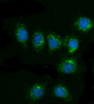
ARG40691 anti-CD81 antibody ICC/IF image
Immunofluorescence: RM-1 cells were blocked with 10% goat serum and then stained with ARG40691 anti-CD81 antibody (green) at 5 µg/ml dilution, overnight at 4°C. DAPI (blue) for nuclear staining.
-
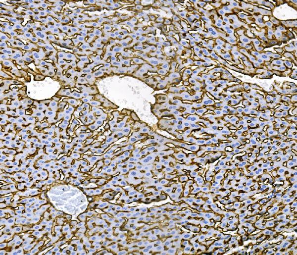
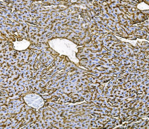
ARG40691 anti-CD81 antibody IHC-P image
Immunohistochemistry: Paraffin-embedded Mouse liver tissue. Antigen Retrieval: Heat mediation was performed in EDTA buffer (pH 8.0). The tissue section was blocked with 10% goat serum. The tissue section was then stained with ARG40691 anti-CD81 antibody at 1 µg/ml dilution, overnight at 4°C.
-
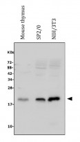
ARG40691 anti-CD81 antibody WB image
Western blot: 30 µg of sample under reducing conditions. Mouse thymus, SP2/0, NIH/3T3 whole cell lysates stained with ARG40691 anti-CD81 antibody at 0.5 µg/ml dilution, overnight at 4°C.
-
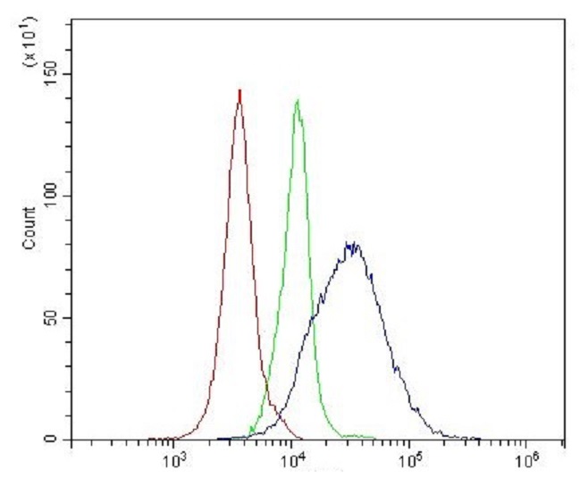
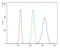
ARG40691 anti-CD81 antibody FACS image
Flow Cytometry: Mouse spleen cells were blocked with 10% normal goat serum and then stained with ARG40691 anti-CD81 antibody (blue) at 1 µg/10^6 cells for 30 min at 20°C, followed by incubation with DyLight®488 labelled secondary antibody. Isotype control antibody (green) was rabbit IgG (1 µg/10^6 cells) used under the same conditions. Unlabelled sample (red) was also used as a control.
-
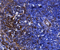
ARG40691 anti-CD81 antibody IHC-P image
Immunohistochemistry: Paraffin-embedded Mouse spleen tissue. Antigen Retrieval: Heat mediation was performed in EDTA buffer (pH 8.0). The tissue section was blocked with 10% goat serum. The tissue section was then stained with ARG40691 anti-CD81 antibody at 1 µg/ml dilution, overnight at 4°C.
-
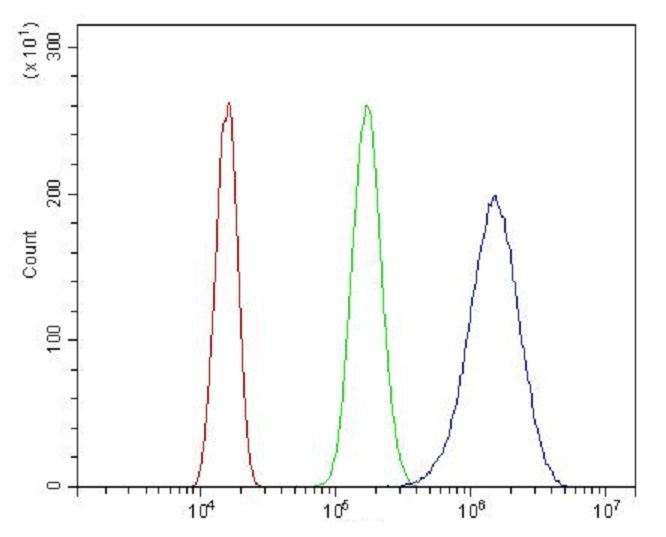
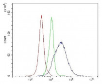
ARG40691 anti-CD81 antibody FACS image
Flow Cytometry: Neuro-2a cells were blocked with 10% normal goat serum and then stained with ARG40691 anti-CD81 antibody (blue) at 1 µg/10^6 cells for 30 min at 20°C, followed by incubation with DyLight®488 labelled secondary antibody. Isotype control antibody (green) was rabbit IgG (1 µg/10^6 cells) used under the same conditions. Unlabelled sample (red) was also used as a control.