ARG65460
anti-CD79a antibody [HM57] (FITC)
anti-CD79a antibody [HM57] (FITC) for Flow cytometry and Human,Mouse,Rat,Bovine,Chicken,Guinea pig,Horse,Opossum,Pig,Rabbit
Cancer antibody; Developmental Biology antibody; Immune System antibody
Overview
| Product Description | FITC-conjugated Mouse Monoclonal antibody [HM57] recognizes CD79a |
|---|---|
| Tested Reactivity | Hu, Ms, Rat, Bov, Chk, Gpig, Hrs, Opossum, Pig, Rb |
| Tested Application | FACS |
| Specificity | The clone HM57 interacts with CD79a (Ig alpha), a 40-45 kDa subunit of B cell antigen-specific receptor (BCR) and its early developmental forms. HLDA V; WS Code BC cB018 HLDA VI; WS Code BP 193 HLDA VI; WS Code BP 89 HLDA VI; WS Code B B103 HLDA VI; WS Code B CD79.4 |
| Host | Mouse |
| Clonality | Monoclonal |
| Clone | HM57 |
| Isotype | IgG1 |
| Target Name | CD79a |
| Antigen Species | Human |
| Immunogen | Synthetic peptide corresponding to amino acids 202-216 of human CD79a_x000D_ |
| Conjugation | FITC |
| Alternate Names | Surface IgM-associated protein; B-cell antigen receptor complex-associated protein alpha chain; Membrane-bound immunoglobulin-associated protein; Ig-alpha; MB-1 membrane glycoprotein; MB-1; IGA; CD antigen CD79a |
Application Instructions
| Application Suggestion |
|
||||
|---|---|---|---|---|---|
| Application Note | * The dilutions indicate recommended starting dilutions and the optimal dilutions or concentrations should be determined by the scientist. |
Properties
| Form | Liquid |
|---|---|
| Purification Note | The purified antibody is conjugated with Fluorescein isothiocyanate (FITC) under optimum conditions. The reagent is free of unconjugated FITC and adjusted for direct use. No reconstitution is necessary. |
| Buffer | PBS, 15 mM Sodium azide and 0.2% (w/v) high-grade protease free BSA |
| Preservative | 15 mM Sodium azide |
| Stabilizer | 0.2% (w/v) high-grade protease free BSA |
| Storage Instruction | Aliquot and store in the dark at 2-8°C. Keep protected from prolonged exposure to light. Avoid repeated freeze/thaw cycles. Suggest spin the vial prior to opening. The antibody solution should be gently mixed before use. |
| Note | For laboratory research only, not for drug, diagnostic or other use. |
Bioinformation
| Database Links | |
|---|---|
| Gene Symbol | CD79A |
| Gene Full Name | CD79a molecule, immunoglobulin-associated alpha |
| Background | CD79a: The B lymphocyte antigen receptor is a multimeric complex that includes the antigen-specific component, surface immunoglobulin (Ig). Surface Ig non-covalently associates with two other proteins, Ig-alpha and Ig-beta, which are necessary for expression and function of the B-cell antigen receptor. This gene encodes the Ig-alpha protein of the B-cell antigen component. Alternatively spliced transcript variants encoding different isoforms have been described. [provided by RefSeq, Jul 2008] |
| Function | CD79a is required in cooperation with CD79b for initiation of the signal transduction cascade activated by binding of antigen to the B-cell antigen receptor complex (BCR) which leads to internalization of the complex, trafficking to late endosomes and antigen presentation. Also required for BCR surface expression and for efficient differentiation of pro- and pre-B-cells. Stimulates SYK autophosphorylation and activation. Binds to BLNK, bringing BLNK into proximity with SYK and allowing SYK to phosphorylate BLNK. Also interacts with and increases activity of some Src-family tyrosine kinases. Represses BCR signaling during development of immature B-cells. [UniProt] |
| Highlight | Related products: CD79a antibodies; Anti-Mouse IgG secondary antibodies; Related news: Tumor-Infiltrating Lymphocytes (TILs) |
| Research Area | Cancer antibody; Developmental Biology antibody; Immune System antibody |
| Calculated MW | 25 kDa |
| PTM | Phosphorylated on tyrosine, serine and threonine residues upon B-cell activation. Phosphorylation of tyrosine residues by Src-family kinases is an early and essential feature of the BCR signaling cascade. The phosphorylated tyrosines serve as docking sites for SH2-domain containing kinases, leading to their activation which in turn leads to phosphorylation of downstream targets. Phosphorylated by LYN. Phosphorylation of serine and threonine residues may prevent subsequent tyrosine phosphorylation. Arginine methylation in the ITAM domain may interfere with the binding of SYK. It promotes signals leading to B-cell differentiation (By similarity). |
Images (3) Click the Picture to Zoom In
-
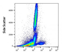
ARG65460 anti-CD79a antibody [HM57] (FITC) FACS image
Flow Cytometry: Human peripheral whole blood stained with ARG65460 anti-CD79a antibody [HM57] (FITC) (4 µl reagent / 100 µl of peripheral whole blood).
-

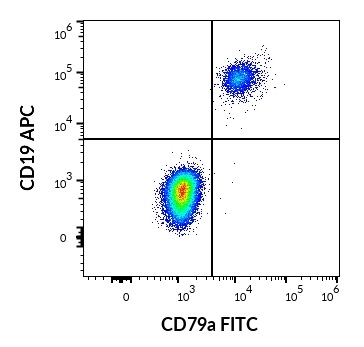
ARG65460 anti-CD79a antibody [HM57] (FITC) FACS image
Flow Cytometry: Human lymphocytes stained with ARG53782 anti-CD19 antibody [LT19] (APC) (10 µl reagent / 100 µl of peripheral whole blood) and ARG65460 anti-CD79a antibody [HM57] (FITC) (4 µl reagent / 100 µl of peripheral whole blood).
-
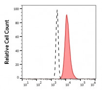
ARG65460 anti-CD79a antibody [HM57] (FITC) FACS image
Flow Cytometry: Separation of human CD79a positive B cells (red-filled) from neutrophil granulocytes (black-dashed). Human peripheral whole blood stained with ARG65460 anti-CD79a antibody [HM57] (FITC) (4 µl reagent / 100 µl of peripheral whole blood).