ARG40207
anti-CD79a antibody
anti-CD79a antibody for IHC-Formalin-fixed paraffin-embedded sections,Western blot and Human,Rat
Overview
| Product Description | Rabbit Polyclonal antibody recognizes CD79a |
|---|---|
| Tested Reactivity | Hu, Rat |
| Tested Application | IHC-P, WB |
| Host | Rabbit |
| Clonality | Polyclonal |
| Isotype | IgG |
| Target Name | CD79a |
| Antigen Species | Human |
| Immunogen | Recombinant protein corresponding to T121-P226 of Human CD79a. |
| Conjugation | Un-conjugated |
| Alternate Names | Surface IgM-associated protein; B-cell antigen receptor complex-associated protein alpha chain; Membrane-bound immunoglobulin-associated protein; Ig-alpha; MB-1 membrane glycoprotein; MB-1; IGA; CD antigen CD79a |
Application Instructions
| Application Suggestion |
|
||||||
|---|---|---|---|---|---|---|---|
| Application Note | IHC-P: Antigen Retrieval: By heat mediation. * The dilutions indicate recommended starting dilutions and the optimal dilutions or concentrations should be determined by the scientist. |
Properties
| Form | Liquid |
|---|---|
| Purification | Affinity purification with immunogen. |
| Buffer | 0.9% NaCl, 0.2% Na2HPO4, 0.05% Sodium azide and 5% BSA. |
| Preservative | 0.05% Sodium azide |
| Stabilizer | 5% BSA |
| Concentration | 0.5 mg/ml |
| Storage Instruction | For continuous use, store undiluted antibody at 2-8°C for up to a week. For long-term storage, aliquot and store at -20°C or below. Storage in frost free freezers is not recommended. Avoid repeated freeze/thaw cycles. Suggest spin the vial prior to opening. The antibody solution should be gently mixed before use. |
| Note | For laboratory research only, not for drug, diagnostic or other use. |
Bioinformation
| Database Links |
Swiss-port # P11912 Human B-cell antigen receptor complex-associated protein alpha chain |
|---|---|
| Gene Symbol | CD79A |
| Gene Full Name | CD79a molecule, immunoglobulin-associated alpha |
| Background | CD79a: The B lymphocyte antigen receptor is a multimeric complex that includes the antigen-specific component, surface immunoglobulin (Ig). Surface Ig non-covalently associates with two other proteins, Ig-alpha and Ig-beta, which are necessary for expression and function of the B-cell antigen receptor. This gene encodes the Ig-alpha protein of the B-cell antigen component. Alternatively spliced transcript variants encoding different isoforms have been described. [provided by RefSeq, Jul 2008] |
| Function | CD79a is required in cooperation with CD79b for initiation of the signal transduction cascade activated by binding of antigen to the B-cell antigen receptor complex (BCR) which leads to internalization of the complex, trafficking to late endosomes and antigen presentation. Also required for BCR surface expression and for efficient differentiation of pro- and pre-B-cells. Stimulates SYK autophosphorylation and activation. Binds to BLNK, bringing BLNK into proximity with SYK and allowing SYK to phosphorylate BLNK. Also interacts with and increases activity of some Src-family tyrosine kinases. Represses BCR signaling during development of immature B-cells. [UniProt] |
| Cellular Localization | Cell membrane; Single-pass type I membrane protein. Note=Following antigen binding, the BCR has been shown to translocate from detergent-soluble regions of the cell membrane to lipid rafts although signal transduction through the complex can also occur outside lipid rafts. [UniProt] |
| Highlight | Related products: CD79a antibodies; Anti-Rabbit IgG secondary antibodies; Related news: Tumor-Infiltrating Lymphocytes (TILs) |
| Calculated MW | 25 kDa |
| PTM | Phosphorylated on tyrosine, serine and threonine residues upon B-cell activation. Phosphorylation of tyrosine residues by Src-family kinases is an early and essential feature of the BCR signaling cascade. The phosphorylated tyrosines serve as docking sites for SH2-domain containing kinases, leading to their activation which in turn leads to phosphorylation of downstream targets. Phosphorylated by LYN. Phosphorylation of serine and threonine residues may prevent subsequent tyrosine phosphorylation. Arginine methylation in the ITAM domain may interfere with the binding of SYK. It promotes signals leading to B-cell differentiation (By similarity). [UniProt] |
Images (3) Click the Picture to Zoom In
-
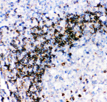
ARG40207 anti-CD79a antibody IHC-P image
Immunohistochemistry: Paraffin-embedded Human tonsil stained with ARG40207 anti-CD79a antibody.
-
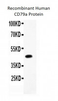
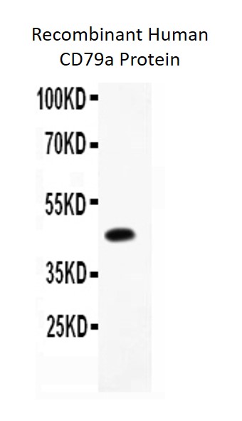
ARG40207 anti-CD79a antibody WB image
Western blot: 0.5 ng of Recombinant Human CD79a Protein stained with ARG40207 anti-CD79a antibody at 0.5 µg/ml dilution.
-
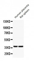
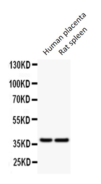
ARG40207 anti-CD79a antibody WB image
Western blot: 50 µg of Human placenta and Rat spleen lysates stained with ARG40207 anti-CD79a antibody at 0.5 µg/ml dilution.