ARG41769
anti-CD71 / Transferrin Receptor antibody
anti-CD71 / Transferrin Receptor antibody for Flow cytometry,ICC/IF,IHC-Frozen sections,IHC-Formalin-fixed paraffin-embedded sections,Western blot and Human
Overview
| Product Description | Rabbit Polyclonal antibody recognizes CD71 / Transferrin Receptor |
|---|---|
| Tested Reactivity | Hu |
| Tested Application | FACS, ICC/IF, IHC-Fr, IHC-P, WB |
| Host | Rabbit |
| Clonality | Polyclonal |
| Isotype | IgG |
| Target Name | CD71 / Transferrin Receptor |
| Antigen Species | Human |
| Immunogen | Recombinant protein corresponding to M1-N198 of Human CD71 / Transferrin Receptor. |
| Conjugation | Un-conjugated |
| Alternate Names | TFR1; CD antigen CD71; CD71; T9; p90; TR; Trfr; Transferrin receptor protein 1; TRFR; sTfR; TfR1; TfR; TFR |
Application Instructions
| Application Suggestion |
|
||||||||||||
|---|---|---|---|---|---|---|---|---|---|---|---|---|---|
| Application Note | IHC-P: Antigen Retrieval: Heat mediation was performed in Citrate buffer (pH 6.0, epitope retrieval solution) for 20 min. * The dilutions indicate recommended starting dilutions and the optimal dilutions or concentrations should be determined by the scientist. |
||||||||||||
| Observed Size | ~ 95 kDa |
Properties
| Form | Liquid |
|---|---|
| Purification | Affinity purification with immunogen. |
| Buffer | 0.2% Na2HPO4, 0.9% NaCl, 0.05% Sodium azide and 5% BSA. |
| Preservative | 0.05% Sodium azide |
| Stabilizer | 5% BSA |
| Concentration | 0.5 mg/ml |
| Storage Instruction | For continuous use, store undiluted antibody at 2-8°C for up to a week. For long-term storage, aliquot and store at -20°C or below. Storage in frost free freezers is not recommended. Avoid repeated freeze/thaw cycles. Suggest spin the vial prior to opening. The antibody solution should be gently mixed before use. |
| Note | For laboratory research only, not for drug, diagnostic or other use. |
Bioinformation
| Database Links | |
|---|---|
| Gene Symbol | TFRC |
| Gene Full Name | transferrin receptor |
| Background | This gene encodes a cell surface receptor necessary for cellular iron uptake by the process of receptor-mediated endocytosis. This receptor is required for erythropoiesis and neurologic development. Multiple alternatively spliced variants have been identified. [provided by RefSeq, Sep 2015] |
| Function | Cellular uptake of iron occurs via receptor-mediated endocytosis of ligand-occupied transferrin receptor into specialized endosomes. Endosomal acidification leads to iron release. The apotransferrin-receptor complex is then recycled to the cell surface with a return to neutral pH and the concomitant loss of affinity of apotransferrin for its receptor. Transferrin receptor is necessary for development of erythrocytes and the nervous system (By similarity). A second ligand, the heditary hemochromatosis protein HFE, competes for binding with transferrin for an overlapping C-terminal binding site. [UniProt] |
| Cellular Localization | Cell membrane; Single-pass type II membrane protein. Melanosome. Note=Identified by mass spectrometry in melanosome fractions from stage I to stage IV. Transferrin receptor protein 1, serum form: Secreted. [UniProt] |
| Calculated MW | 85 kDa |
| PTM | N- and O-glycosylated, phosphorylated and palmitoylated. The serum form is only glycosylated. Proteolytically cleaved on Arg-100 to produce the soluble serum form (sTfR). Palmitoylated on both Cys-62 and Cys-67. Cys-62 seems to be the major site of palmitoylation. [UniProt] |
Images (6) Click the Picture to Zoom In
-
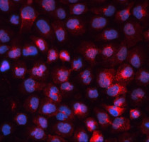
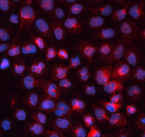
ARG41769 anti-CD71 / Transferrin Receptor antibody ICC/IF image
Immunofluorescence: A431 cells were blocked with 10% goat serum and then stained with ARG41769 anti-CD71 / Transferrin Receptor antibody (red) at 2 µg/ml dilution, overnight at 4°C. DAPI (blue) for nuclear staining.
-
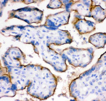
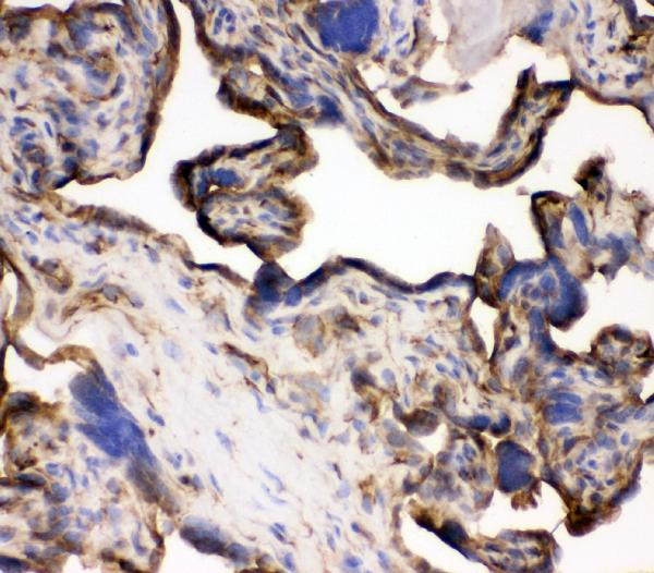
ARG41769 anti-CD71 / Transferrin Receptor antibody IHC-P image
Immunohistochemistry: Paraffin-embedded Human placenta tissue. Antigen Retrieval: Heat mediation was performed in Citrate buffer (pH 6.0, epitope retrieval solution) for 20 min. The tissue section was blocked with 10% goat serum. The tissue section was then stained with ARG41769 anti-CD71 / Transferrin Receptor antibody at 1 µg/ml dilution, overnight at 4°C.
-

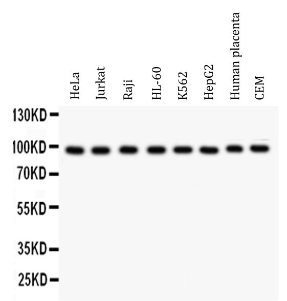
ARG41769 anti-CD71 / Transferrin Receptor antibody WB image
Western blot: 50 µg of samples under reducing conditions. HeLa, Jurkat, Raji, HL-60, K562, HepG2, Human placenta and CEM whole cell lysates stained with ARG41769 anti-CD71 / Transferrin Receptor antibody at 0.5 µg/ml dilution, overnight at 4°C.
-
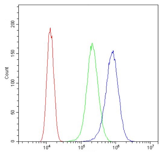
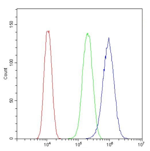
ARG41769 anti-CD71 / Transferrin Receptor antibody FACS image
Flow Cytometry: SiHa cells were blocked with 10% normal goat serum and then stained with ARG41769 anti-CD71 / Transferrin Receptor antibody (blue) at 1 µg/10^6 cells for 30 min at 20°C, followed by incubation with DyLight®488 labelled secondary antibody. Isotype control antibody (green) was Rabbit IgG (1 µg/10^6 cells) used under the same conditions. Unlabelled sample (red) was also used as a control.
-

ARG41769 anti-CD71 / Transferrin Receptor antibody IHC-Fr image
Immunohistochemistry: Frozen section of Human placenta tissue. The tissue section was blocked with 10% goat serum. The tissue section was then stained with ARG41769 anti-CD71 / Transferrin Receptor antibody at 1 µg/ml dilution, overnight at 4°C.
-
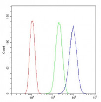
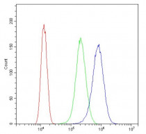
ARG41769 anti-CD71 / Transferrin Receptor antibody FACS image
Flow Cytometry: U87 cells were blocked with 10% normal goat serum and then stained with ARG41769 anti-CD71 / Transferrin Receptor antibody (blue) at 1 µg/10^6 cells for 30 min at 20°C, followed by incubation with DyLight®488 labelled secondary antibody. Isotype control antibody (green) was Rabbit IgG (1 µg/10^6 cells) used under the same conditions. Unlabelled sample (red) was also used as a control.