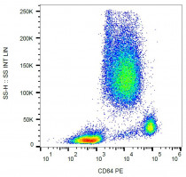
ARG54186
anti-CD64 antibody [10.1] (PE)
anti-CD64 antibody [10.1] (PE) for Flow cytometry and Human,Primates
Immune System antibody
Overview
| Product Description | PE-conjugated Mouse Monoclonal antibody [10.1] recognizes CD64 |
|---|---|
| Tested Reactivity | Hu, NHuPrm |
| Tested Application | FACS |
| Specificity | The clone 10.1 recognizes alpha subunit of CD64/FcgammaRI, a 72 kDa single chain type I glycoprotein, that is expressed on monocytes/macrophages, dendritic cells, and activated granulocytes. HLDA III; WS Code M-250 |
| Host | Mouse |
| Clonality | Monoclonal |
| Clone | 10.1 |
| Isotype | IgG1 |
| Target Name | CD64 |
| Antigen Species | Human |
| Immunogen | Rheumatoid synovial fluid cells and fibronectin purified human monocytes |
| Conjugation | PE |
| Alternate Names | High affinity immunoglobulin gamma Fc receptor I; CD64; Fc-gamma RIA; CD antigen CD64; FcgammaRIa; FCRI; IgG Fc receptor I; CD64A; Fc-gamma RI; FcRI; IGFR1 |
Application Instructions
| Application Suggestion |
|
||||
|---|---|---|---|---|---|
| Application Note | * The dilutions indicate recommended starting dilutions and the optimal dilutions or concentrations should be determined by the scientist. |
Properties
| Form | Liquid |
|---|---|
| Purification Note | The purified antibody is conjugated with R-Phycoerythrin (PE) under optimum conditions. The conjugate is purified by size-exclusion chromatography and adjusted for direct use. No reconstitution is necessary. |
| Buffer | PBS, 15 mM Sodium azide and 0.2% (w/v) high-grade protease free BSA |
| Preservative | 15 mM Sodium azide |
| Stabilizer | 0.2% (w/v) high-grade protease free BSA |
| Storage Instruction | Aliquot and store in the dark at 2-8°C. Keep protected from prolonged exposure to light. Avoid repeated freeze/thaw cycles. Suggest spin the vial prior to opening. The antibody solution should be gently mixed before use. |
| Note | For laboratory research only, not for drug, diagnostic or other use. |
Bioinformation
| Database Links |
Swiss-port # P12314 Human High affinity immunoglobulin gamma Fc receptor I |
|---|---|
| Gene Symbol | FCGR1A |
| Gene Full Name | Fc fragment of IgG, high affinity Ia, receptor (CD64) |
| Background | CD64 (FcgammaRI) is a cell surface receptor for Fc region of IgG. It is composed of specific ligand binding alpha subunit and promiscuous gamma subunit, which is indispensable for tyrosine-based signaling. However, even the alpha subunit can transduce signals leading to cellular effector functions. The isoform FcgammaRIa1 binds human IgG with high affinity, has limited myeloid cell distribution, and a relatively large intracellular domain. Products of related genes include FcgammaRIb and FcgammaRIc isoforms, but these specify low affinity IgG receptors if functionally expressed at all. Besides a role in antigen clearance, FcgammaRI (a1) can potently enhance MHC class I and II antigen presentation in vitro and in vivo. |
| Function | High affinity receptor for the Fc region of immunoglobulins gamma. Functions in both innate and adaptive immune responses. [UniProt] |
| Research Area | Immune System antibody |
| Calculated MW | 43 kDa |
| PTM | Phosphorylated on serine residues. |
Images (1) Click the Picture to Zoom In
Clone References


