ARG67138
anti-CD63 antibody
anti-CD63 antibody for ICC/IF,IHC-Formalin-fixed paraffin-embedded sections,Western blot and Human,Mouse,Rat
Overview
| Product Description | Rabbit Polyclonal antibody recognizes CD63 |
|---|---|
| Tested Reactivity | Hu, Ms, Rat |
| Tested Application | ICC/IF, IHC-P, WB |
| Host | Rabbit |
| Clonality | Polyclonal |
| Isotype | IgG |
| Target Name | CD63 |
| Antigen Species | Human |
| Immunogen | Synthetic peptide of Human CD63. |
| Conjugation | Un-conjugated |
| Alternate Names | CD63; CD63 Molecule; TSPAN30; ME491; MLA1; Ocular Melanoma-Associated Antigen; CD63 Antigen (Melanoma 1 Antigen); Melanoma-Associated Antigen ME491; Tetraspanin-30; Granulophysin; CD63 Antigen; Tspan-30; OMA81H; Limp1; Lysosomal-Associated Membrane Protein 3; Lysosome Integral Membrane Protein 1; Melanoma-Associated Antigen MLA1; LAMP-3 |
Application Instructions
| Application Suggestion |
|
||||||||
|---|---|---|---|---|---|---|---|---|---|
| Application Note | * The dilutions indicate recommended starting dilutions and the optimal dilutions or concentrations should be determined by the scientist. |
Properties
| Form | Liquid |
|---|---|
| Purification | Affinity chromatography purified |
| Buffer | PBS, 0.02% Sodium azide, 0.5% BSA and 50% Glycerol |
| Preservative | 0.02% Sodium azide |
| Stabilizer | 0.5% BSA, 50% Glycerol |
| Concentration | 1 mg/ml |
| Storage Instruction | For continuous use, store undiluted antibody at 2-8°C for up to a week. For long-term storage, aliquot and store at -20°C or below. Storage in frost free freezers is not recommended. Avoid repeated freeze/thaw cycles. Suggest spin the vial prior to opening. The antibody solution should be gently mixed before use. |
| Note | For laboratory research only, not for drug, diagnostic or other use. |
Bioinformation
| Database Links | |
|---|---|
| Gene Symbol | CD63 |
| Gene Full Name | CD63 Molecule |
| Background | The protein encoded by this gene is a member of the transmembrane 4 superfamily, also known as the tetraspanin family. Most of these members are cell-surface proteins that are characterized by the presence of four hydrophobic domains. The proteins mediate signal transduction events that play a role in the regulation of cell development, activation, growth and motility. The encoded protein is a cell surface glycoprotein that is known to complex with integrins. It may function as a blood platelet activation marker. Deficiency of this protein is associated with Hermansky-Pudlak syndrome. Also this gene has been associated with tumor progression. Alternative splicing results in multiple transcript variants encoding different protein isoforms. [provided by RefSeq, Apr 2012] |
| Function | Functions as a cell surface receptor for TIMP1 and plays a role in the activation of cellular signaling cascades. Plays a role in the activation of ITGB1 and integrin signaling, leading to the activation of AKT, FAK/PTK2 and MAP kinases. Promotes cell survival, reorganization of the actin cytoskeleton, cell adhesion, spreading and migration, via its role in the activation of AKT and FAK/PTK2. Plays a role in VEGFA signaling via its role in regulating the internalization of KDR/VEGFR2. Plays a role in intracellular vesicular transport processes, and is required for normal trafficking of the PMEL luminal domain that is essential for the development and maturation of melanocytes. Plays a role in the adhesion of leukocytes onto endothelial cells via its role in the regulation of SELP trafficking. May play a role in mast cell degranulation in response to Ms4a2/FceRI stimulation, but not in mast cell degranulation in response to other stimuli. [Uniprot] |
| Cellular Localization | Cell membrane, Endosome, Lysosome, Membrane, Secreted. [Uniprot] |
| Calculated MW | 26 kDa |
| PTM | Glycoprotein, Lipoprotein, Palmitate. [Uniprot] |
Images (3) Click the Picture to Zoom In
-

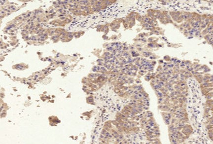
ARG67138 anti-CD63 antibody IHC-P image
Immunohistochemistry: Human liver cancer stained with ARG67138 anti-CD63 antibody at 1:200 dilution.
-
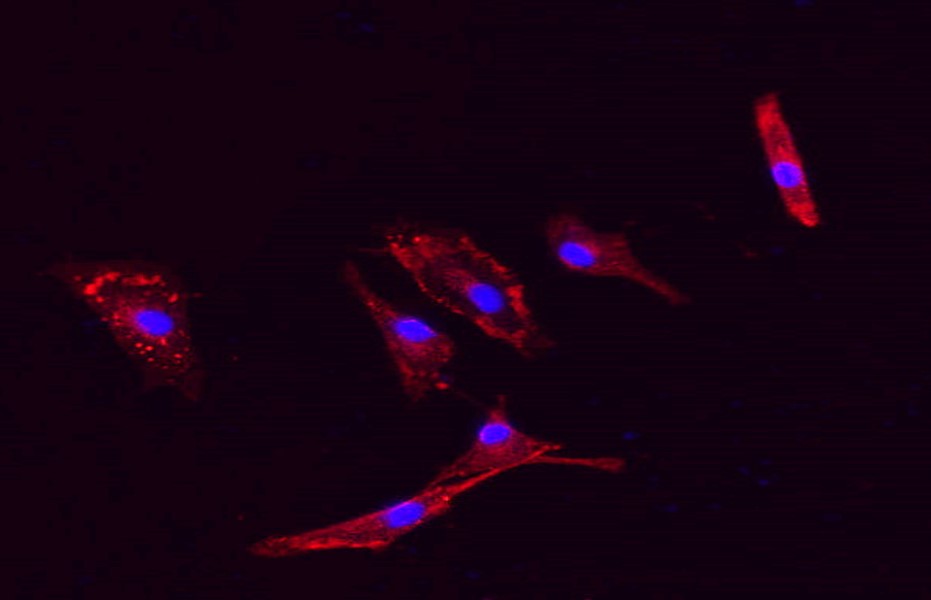
ARG67138 anti-CD63 antibody ICC/IF image
Immunofluorescence: A549 stained with ARG67138 anti-CD63 antibody at 1:200 dilution.
-
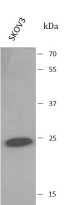
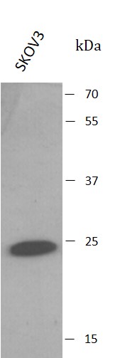
ARG67138 anti-CD63 antibody WB image
Western blot: SKOV3 stained with ARG67138 anti-CD63 antibody.