ARG42580
anti-CD54 / ICAM1 antibody
anti-CD54 / ICAM1 antibody for Flow cytometry,ICC/IF,IHC-Formalin-fixed paraffin-embedded sections,Western blot and Human,Mouse,Rat
Overview
| Product Description | Rabbit Polyclonal antibody recognizes CD54 / ICAM1 |
|---|---|
| Tested Reactivity | Hu, Ms, Rat |
| Tested Application | FACS, ICC/IF, IHC-P, WB |
| Host | Rabbit |
| Clonality | Polyclonal |
| Isotype | IgG |
| Target Name | CD54 / ICAM1 |
| Antigen Species | Human |
| Immunogen | Recombinant protein corresponding to Q28-R268 of Human CD54 / ICAM1. |
| Conjugation | Un-conjugated |
| Alternate Names | CD54; CD antigen CD54; BB2; P3.58; Intercellular adhesion molecule 1; Major group rhinovirus receptor; ICAM-1 |
Application Instructions
| Application Suggestion |
|
||||||||||
|---|---|---|---|---|---|---|---|---|---|---|---|
| Application Note | IHC-P: Antigen Retrieval: Heat mediation was performed in EDTA buffer (pH 8.0). * The dilutions indicate recommended starting dilutions and the optimal dilutions or concentrations should be determined by the scientist. |
||||||||||
| Observed Size | ~ 95 kDa |
Properties
| Form | Liquid |
|---|---|
| Purification | Affinity purification with immunogen. |
| Buffer | 0.2% Na2HPO4, 0.9% NaCl, 0.05% Sodium azide and 4% Trehalose. |
| Preservative | 0.05% Sodium azide |
| Stabilizer | 4% Trehalose |
| Concentration | 0.5 mg/ml |
| Storage Instruction | For continuous use, store undiluted antibody at 2-8°C for up to a week. For long-term storage, aliquot and store at -20°C or below. Storage in frost free freezers is not recommended. Avoid repeated freeze/thaw cycles. Suggest spin the vial prior to opening. The antibody solution should be gently mixed before use. |
| Note | For laboratory research only, not for drug, diagnostic or other use. |
Bioinformation
| Database Links | |
|---|---|
| Gene Symbol | ICAM1 |
| Gene Full Name | intercellular adhesion molecule 1 |
| Background | This gene encodes a cell surface glycoprotein which is typically expressed on endothelial cells and cells of the immune system. It binds to integrins of type CD11a / CD18, or CD11b / CD18 and is also exploited by Rhinovirus as a receptor. [provided by RefSeq, Jul 2008] |
| Function | ICAM proteins are ligands for the leukocyte adhesion protein LFA-1 (integrin alpha-L/beta-2). During leukocyte trans-endothelial migration, ICAM1 engagement promotes the assembly of endothelial apical cups through ARHGEF26/SGEF and RHOG activation. (Microbial infection) Acts as a receptor for major receptor group rhinovirus A-B capsid proteins. (Microbial infection) Acts as a receptor for Coxsackievirus A21 capsid proteins. (Microbial infection) Upon Kaposi's sarcoma-associated herpesvirus/HHV-8 infection, is degraded by viral E3 ubiquitin ligase MIR2, presumably to prevent lysis of infected cells by cytotoxic T-lymphocytes and NK cell. [UniProt] |
| Cellular Localization | Membrane; Single-pass type I membrane protein. [UniProt] |
| Calculated MW | 58 kDa |
| PTM | Monoubiquitinated, which is promoted by MARCH9 and leads to endocytosis. [UniProt] |
Images (6) Click the Picture to Zoom In
-
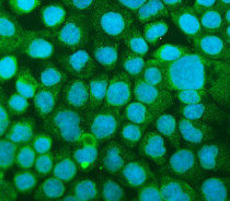
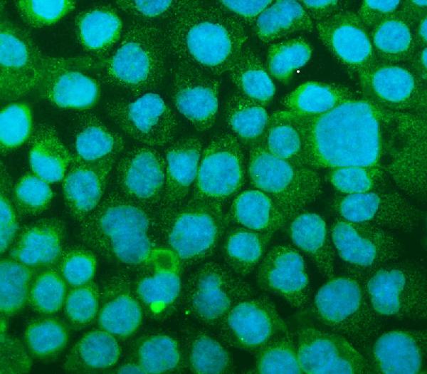
ARG42580 anti-CD54 / ICAM1 antibody ICC/IF image
Immunofluorescence: A431 cells were blocked with 10% goat serum and then stained with ARG42580 anti-CD54 / ICAM1 antibody (green) at 2 µg/ml dilution, overnight at 4°C. DAPI (blue) for nuclear staining.
-

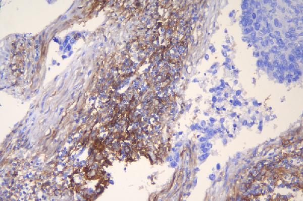
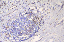
ARG42580 anti-CD54 / ICAM1 antibody IHC-P image
Immunohistochemistry: Paraffin-embedded Human lung cancer tissue. Antigen Retrieval: Heat mediation was performed in EDTA buffer (pH 8.0). The tissue section was blocked with 10% goat serum. The tissue section was then stained with ARG42580 anti-CD54 / ICAM1 antibody at 1 µg/ml dilution, overnight at 4°C.
-
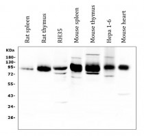
ARG42580 anti-CD54 / ICAM1 antibody WB image
Western blot: 50 µg of samples under reducing conditions. Rat spleen, Rat thymus, RH35, Mouse spleen, Mouse thymus, Hepa 1-6 and Mouse heart lysates stained with ARG42580 anti-CD54 / ICAM1 antibody at 0.5 µg/ml dilution, overnight at 4°C.
-
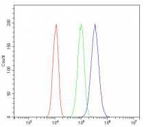
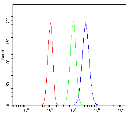
ARG42580 anti-CD54 / ICAM1 antibody FACS image
Flow Cytometry: PC-3 cells were blocked with 10% normal goat serum and then stained with ARG42580 anti-CD54 / ICAM1 antibody (blue) at 1 µg/10^6 cells for 30 min at 20°C, followed by incubation with DyLight®488 labelled secondary antibody. Isotype control antibody (green) was Rabbit IgG (1 µg/10^6 cells) used under the same conditions. Unlabelled sample (red) was also used as a control.
-
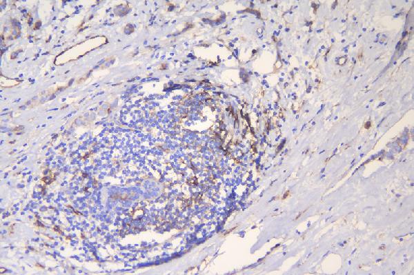
ARG42580 anti-CD54 / ICAM1 antibody IHC-P image
Immunohistochemistry: Paraffin-embedded Human mammary cancer tissue. Antigen Retrieval: Heat mediation was performed in EDTA buffer (pH 8.0). The tissue section was blocked with 10% goat serum. The tissue section was then stained with ARG42580 anti-CD54 / ICAM1 antibody at 1 µg/ml dilution, overnight at 4°C.
-
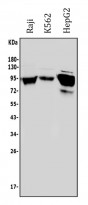
ARG42580 anti-CD54 / ICAM1 antibody WB image
Western blot: 50 µg of samples under reducing conditions. Raji, K562 and HepG2 whole cell lysates stained with ARG42580 anti-CD54 / ICAM1 antibody at 0.5 µg/ml dilution, overnight at 4°C.