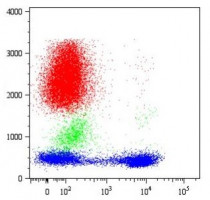
ARG62877
anti-CD5 antibody [CRIS1]
anti-CD5 antibody [CRIS1] for ELISA,Flow cytometry,IHC-Frozen sections,Immunoprecipitation,Western blot and Human
Developmental Biology antibody; Immune System antibody
Overview
| Product Description | Mouse Monoclonal antibody [CRIS1] recognizes CD5 |
|---|---|
| Tested Reactivity | Hu |
| Tested Application | ELISA, FACS, IHC-Fr, IP, WB |
| Specificity | The clone CRIS1 reacts with the cell surface glycoprotein CD5, a 67kDa single-chain transmembrane glycoprotein expressed on mature T lymphocytes, most of thymocytes and B lymphocytes subset (B-1a lymphocytes). HLDA I; WS Code T 29 HLDA III; WS Code T 530 |
| Host | Mouse |
| Clonality | Monoclonal |
| Clone | CRIS1 |
| Isotype | IgG2a |
| Target Name | CD5 |
| Antigen Species | Human |
| Immunogen | stimulated human leukocytes |
| Conjugation | Un-conjugated |
| Alternate Names | CD antigen CD5; Lymphocyte antigen T1/Leu-1; LEU1; T-cell surface glycoprotein CD5; T1 |
Application Instructions
| Application Suggestion |
|
||||||||||||
|---|---|---|---|---|---|---|---|---|---|---|---|---|---|
| Application Note | ELISA: The antibody CRIS1 can be used in the Sandwich ELISA as the detection antibody in pair with the capture antibody MEM-32 * The dilutions indicate recommended starting dilutions and the optimal dilutions or concentrations should be determined by the scientist. |
||||||||||||
| Positive Control | FACS: Peripheral Blood Lymphocytes (PBL), Jurkat human leukemia T-cell line, HPB human leukemia T-cell line and MOLT-4 human leukemia T-cell line. WB: Jurkat human leukemia T-cell line and HPB human leukemia T-cell line. |
Properties
| Form | Liquid |
|---|---|
| Purification | Purified from hybridoma culture supernatant by protein-A affinity chromatography. |
| Purity | > 95% (by SDS-PAGE) |
| Buffer | PBS (pH 7.4) and 15 mM Sodium azide |
| Preservative | 15 mM Sodium azide |
| Concentration | 1 mg/ml |
| Storage Instruction | For continuous use, store undiluted antibody at 2-8°C for up to a week. For long-term storage, aliquot and store at -20°C or below. Storage in frost free freezers is not recommended. Avoid repeated freeze/thaw cycles. Suggest spin the vial prior to opening. The antibody solution should be gently mixed before use. |
| Note | For laboratory research only, not for drug, diagnostic or other use. |
Bioinformation
| Database Links | |
|---|---|
| Gene Symbol | CD5 |
| Gene Full Name | CD5 molecule |
| Background | CD5 antigen (T1; 67 kDa) is a human cell surface T-lymphocyte single-chain transmembrane glycoprotein. CD5 is expressed on all mature T-lymphocytes, most of thymocytes, subset of B-lymphocytes and on many T-cell leukemias and lymphomas. It is a type I membrane glycoprotein whose extracellular region contains three scavenger receptor cysteine-rich (SRCR) domains. The CD5 is a signal transducing molecule whose cytoplasmic tail is devoid of any intrinsic catalytic activity. CD5 modulates signaling through the antigen-specific receptor complex (TCR and BCR). CD5 crosslinking induces extracellular Ca++ mobilization, tyrosine phosphorylation of intracellular proteins and DAG production. Preliminary evidence shows protein associations with ZAP-70, p56lck, p59fyn, PC-PLC, etc. CD5 may serve as a dual receptor, giving either stimulatory or inhibitory signals depending both on the cell type and development stage. In thymocytes and B1a cells seems to provide inhibitory signals, in peripheral mature T lymhocytes it acts as a costimulatory signal receptor. CD5 is the phenotypic marker of a B cell subpopulation involved in the production of autoreactive antibodies. Disease relevance: CD5 is a phenotypic marker for some B cell lymphoproliferative disorders (B-CLL, Hairy cell leukemia, etc.). The CD5+ popuation is expanded in some autoimmune disorders (Rheumatoid Arthritis, etc.). Herpes virus infections induce loss of CD5 expression in the expanded CD8+ human T cells. |
| Function | May act as a receptor in regulating T-cell proliferation. [UniProt] |
| Research Area | Developmental Biology antibody; Immune System antibody |
| Calculated MW | 55 kDa |
| PTM | Phosphorylated on tyrosine residues by LYN; this creates binding sites for PTPN6/SHP-1. |
Images (1) Click the Picture to Zoom In
Clone References


