ARG40482
anti-CD46 antibody
anti-CD46 antibody for Flow cytometry,ICC/IF,IHC-Formalin-fixed paraffin-embedded sections,Western blot and Human,Mouse,Rat
Overview
| Product Description | Rabbit Polyclonal antibody recognizes CD46 |
|---|---|
| Tested Reactivity | Hu, Ms, Rat |
| Tested Application | FACS, ICC/IF, IHC-P, WB |
| Host | Rabbit |
| Clonality | Polyclonal |
| Isotype | IgG |
| Target Name | CD46 |
| Antigen Species | Human |
| Immunogen | Synthetic peptide corresponding to aa. 366-392 of Human CD46. (YRYLQRRKKKGTYLTDETHREVKFTSL) |
| Conjugation | Un-conjugated |
| Alternate Names | MIC10; TLX; CD antigen CD46; Trophoblast leukocyte common antigen; AHUS2; TRA2.10; Membrane cofactor protein; MCP |
Application Instructions
| Application Suggestion |
|
||||||||||
|---|---|---|---|---|---|---|---|---|---|---|---|
| Application Note | IHC-P: Antigen Retrieval: Heat mediation was performed in Citrate buffer (pH 6.0) for 20 min. * The dilutions indicate recommended starting dilutions and the optimal dilutions or concentrations should be determined by the scientist. |
Properties
| Form | Liquid |
|---|---|
| Purification | Affinity purification with immunogen. |
| Buffer | 0.2% Na2HPO4, 0.9% NaCl, 0.05% Sodium azide and 5% BSA. |
| Preservative | 0.05% Sodium azide |
| Stabilizer | 5% BSA |
| Concentration | 0.5 mg/ml |
| Storage Instruction | For continuous use, store undiluted antibody at 2-8°C for up to a week. For long-term storage, aliquot and store at -20°C or below. Storage in frost free freezers is not recommended. Avoid repeated freeze/thaw cycles. Suggest spin the vial prior to opening. The antibody solution should be gently mixed before use. |
| Note | For laboratory research only, not for drug, diagnostic or other use. |
Bioinformation
| Database Links | |
|---|---|
| Gene Symbol | CD46 |
| Gene Full Name | CD46 molecule, complement regulatory protein |
| Background | The protein encoded by this gene is a type I membrane protein and is a regulatory part of the complement system. The encoded protein has cofactor activity for inactivation of complement components C3b and C4b by serum factor I, which protects the host cell from damage by complement. In addition, the encoded protein can act as a receptor for the Edmonston strain of measles virus, human herpesvirus-6, and type IV pili of pathogenic Neisseria. Finally, the protein encoded by this gene may be involved in the fusion of the spermatozoa with the oocyte during fertilization. Mutations at this locus have been associated with susceptibility to hemolytic uremic syndrome. Alternatively spliced transcript variants encoding different isoforms have been described. [provided by RefSeq, Jun 2010] |
| Function | Acts as a cofactor for complement factor I, a serine protease which protects autologous cells against complement-mediated injury by cleaving C3b and C4b deposited on host tissue. May be involved in the fusion of the spermatozoa with the oocyte during fertilization. Also acts as a costimulatory factor for T-cells which induces the differentiation of CD4+ into T-regulatory 1 cells. T-regulatory 1 cells suppress immune responses by secreting interleukin-10, and therefore are thought to prevent autoimmunity. A number of viral and bacterial pathogens seem to exploit this property and directly induce an immunosuppressive phenotype in T-cells by binding to CD46. [UniProt] |
| Cellular Localization | Cytoplasmic vesicle, secretory vesicle, acrosome inner membrane; Single-pass type I membrane protein. Note=Inner acrosomal membrane of spermatozoa. Internalized upon binding of Measles virus, Herpesvirus 6 or Neisseria gonorrhoeae, which results in an increased susceptibility of infected cells to complement-mediated injury. In cancer cells or cells infected by Neisseria, shedding leads to a soluble peptide. [UniProt] |
| Calculated MW | 44 kDa |
| PTM | N-glycosylated on Asn-83; Asn-114 and Asn-273 in most tissues, but probably less N-glycosylated in testis. N-glycosylation on Asn-114 and Asn-273 is required for cytoprotective function. N-glycosylation on Asn-114 is required for Measles virus binding. N-glycosylation on Asn-273 is required for Neisseria binding. N-glycosylation is not required for human adenovirus binding. Extensively O-glycosylated in the Ser/Thr-rich domain. O-glycosylation is required for Neisseria binding but not for Measles virus or human adenovirus binding. In epithelial cells, isoforms B/D/F/H/J/L/3 are phosphorylated by YES1 in response to infection by Neisseria gonorrhoeae; which promotes infectivity. In T-cells, these isoforms may be phosphorylated by LCK. [UniProt] |
Images (6) Click the Picture to Zoom In
-
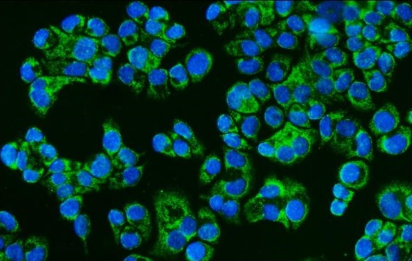
ARG40482 anti-CD46 antibody ICC/IF image
Immunofluorescence: T-47D cells were blocked with 10% goat serum and then stained with ARG40482 anti-CD46 antibody (green) at 5 µg/ml dilution, overnight at 4°C. DAPI (blue) for nuclear staining.
-
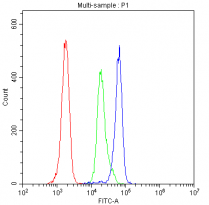
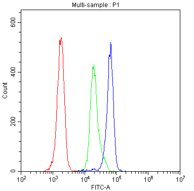
ARG40482 anti-CD46 antibody FACS image
Flow Cytometry: H-PBMC cells were blocked with 10% normal goat serum and then stained with ARG40482 anti-CD46 antibody (blue) at 1 µg/10^6 cells for 30 min at 20°C, followed by incubation with DyLight®488 labelled secondary antibody. Isotype control antibody (green) was Rabbit IgG (1 µg/10^6) used under the same conditions. Unlabelled sample (red) was also used as a control.
-
ARG40482 anti-CD46 antibody IHC-P image
Immunohistochemistry: Paraffin-embedded Human prostatic cancer tissue. Antigen Retrieval: Heat mediation was performed in Citrate buffer (pH 6.0, epitope retrieval solution) for 20 min. The tissue section was blocked with 10% goat serum. The tissue section was then stained with ARG40482 anti-CD46 antibody at 1 µg/ml, overnight at 4°C.
-
ARG40482 anti-CD46 antibody WB image
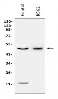
Western blot: 50 µg of sample under reducing conditions. HepG2 and K562 whole cell lysates stained with ARG40482 anti-CD46 antibody at 0.5 µg/ml dilution, overnight at 4°C.
-

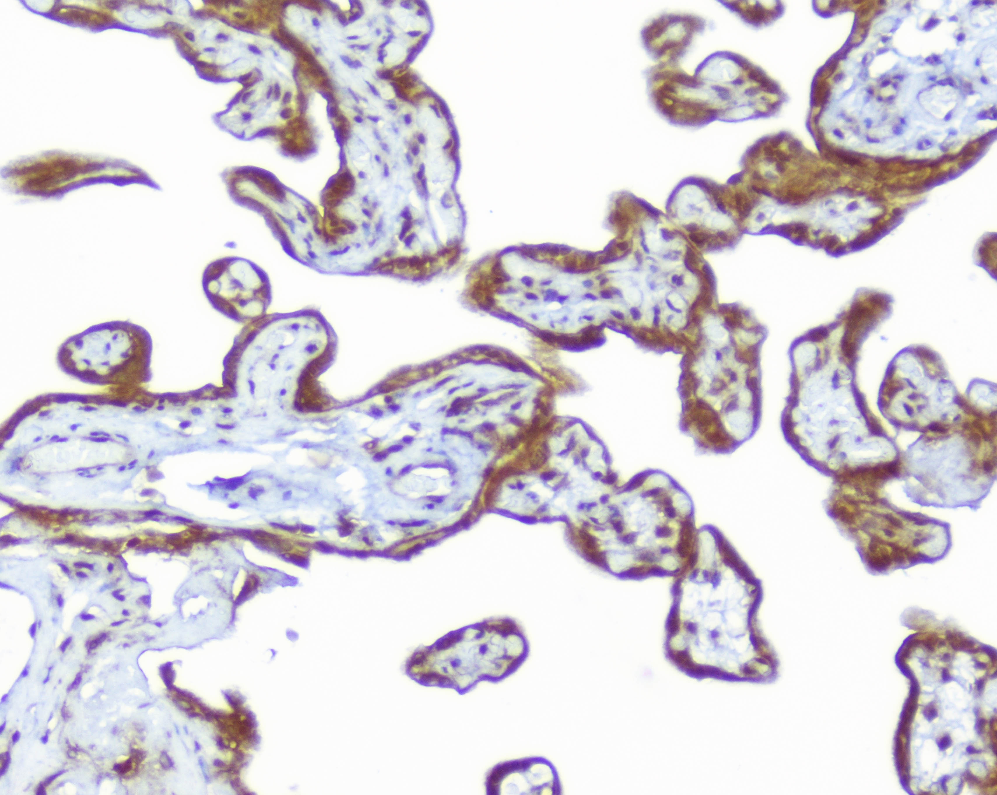
ARG40482 anti-CD46 antibody IHC-P image
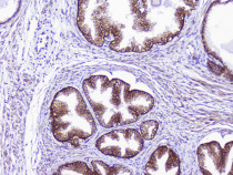
Immunohistochemistry: Paraffin-embedded Human placenta tissue. Antigen Retrieval: Heat mediation was performed in Citrate buffer (pH 6.0, epitope retrieval solution) for 20 min. The tissue section was blocked with 10% goat serum. The tissue section was then stained with ARG40482 anti-CD46 antibody at 1 µg/ml, overnight at 4°C.
-
ARG40482 anti-CD46 antibody WB image
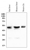
Western blot: 50 µg of sample under reducing conditions. Rat liver, Mouse liver and Neuro-2a whole cell lysates stained with ARG40482 anti-CD46 antibody at 0.5 µg/ml dilution, overnight at 4°C.