ARG66773
anti-CD44 antibody [SQab20206]
anti-CD44 antibody [SQab20206] for IHC-Formalin-fixed paraffin-embedded sections and Human
Overview
| Product Description | Recombinant Rabbit Monoclonal antibody [SQab20206] recognizes CD44 |
|---|---|
| Tested Reactivity | Hu |
| Tested Application | IHC-P |
| Host | Rabbit |
| Clonality | Monoclonal |
| Clone | SQab20206 |
| Isotype | IgG |
| Target Name | CD44 |
| Antigen Species | Human |
| Immunogen | Synthetic peptide within aa. 150-250 of Human CD44. |
| Conjugation | Un-conjugated |
| Alternate Names | MDU2; MDU3; GP90 lymphocyte homing/adhesion receptor; Hermes antigen; Extracellular matrix receptor III; PGP-I; Epican; CDW44; Phagocytic glycoprotein 1; Pgp1; HUTCH-I; MC56; Hyaluronate receptor; CD antigen CD44; Heparan sulfate proteoglycan; CD44 antigen; LHR; IN; HCELL; Phagocytic glycoprotein I; PGP-1; CSPG8; MIC4; ECMR-III; CDw44 |
Application Instructions
| Application Suggestion |
|
||||
|---|---|---|---|---|---|
| Application Note | IHC-P: Antigen Retrieval: Heat mediation was performed in Tris/EDTA buffer (pH 9.0). * The dilutions indicate recommended starting dilutions and the optimal dilutions or concentrations should be determined by the scientist. |
||||
| Positive Control | Tonsil tissue. |
Properties
| Form | Liquid |
|---|---|
| Purification | Purification with Protein A. |
| Buffer | PBS, 0.01% Sodium azide, 40% Glycerol and 0.05% BSA. |
| Preservative | 0.01% Sodium azide |
| Stabilizer | 40% Glycerol and 0.05% BSA |
| Storage Instruction | For continuous use, store undiluted antibody at 2-8°C for up to a week. For long-term storage, aliquot and store at -20°C. Storage in frost free freezers is not recommended. Avoid repeated freeze/thaw cycles. Suggest spin the vial prior to opening. The antibody solution should be gently mixed before use. |
| Note | For laboratory research only, not for drug, diagnostic or other use. |
Bioinformation
| Database Links | |
|---|---|
| Gene Symbol | CD44 |
| Gene Full Name | CD44 molecule (Indian blood group) |
| Background | The protein encoded by this gene is a cell-surface glycoprotein involved in cell-cell interactions, cell adhesion and migration. It is a receptor for hyaluronic acid (HA) and can also interact with other ligands, such as osteopontin, collagens, and matrix metalloproteinases (MMPs). This protein participates in a wide variety of cellular functions including lymphocyte activation, recirculation and homing, hematopoiesis, and tumor metastasis. Transcripts for this gene undergo complex alternative splicing that results in many functionally distinct isoforms, however, the full length nature of some of these variants has not been determined. Alternative splicing is the basis for the structural and functional diversity of this protein, and may be related to tumor metastasis. [provided by RefSeq, Jul 2008] |
| Function | Cell-surface receptor that plays a role in cell-cell interactions, cell adhesion and migration, helping them to sense and respond to changes in the tissue microenvironment (PubMed:16541107, PubMed:19703720, PubMed:22726066). Participates thereby in a wide variety of cellular functions including the activation, recirculation and homing of T-lymphocytes, hematopoiesis, inflammation and response to bacterial infection (PubMed:7528188). Engages, through its ectodomain, extracellular matrix components such as hyaluronan/HA, collagen, growth factors, cytokines or proteases and serves as a platform for signal transduction by assembling, via its cytoplasmic domain, protein complexes containing receptor kinases and membrane proteases (PubMed:18757307, PubMed:23589287). Such effectors include PKN2, the RhoGTPases RAC1 and RHOA, Rho-kinases and phospholipase C that coordinate signaling pathways promoting calcium mobilization and actin-mediated cytoskeleton reorganization essential for cell migration and adhesion (PubMed:15123640). [UniProt] |
| Cellular Localization | Cell membrane; Single-pass type I membrane protein. Cell projection, microvillus. Note=Colocalizes with actin in membrane protrusions at wounding edges. Co-localizes with RDX, EZR and MSN in microvilli. [UniProt] |
| Calculated MW | 82 kDa |
| PTM | Proteolytically cleaved in the extracellular matrix by specific proteinases (possibly MMPs) in several cell lines and tumors. N- and O-glycosylated. O-glycosylation contains more-or-less-sulfated chondroitin sulfate glycans, whose number may affect the accessibility of specific proteinases to their cleavage site(s). It is uncertain if O-glycosylation occurs on Thr-637 or Thr-638. Phosphorylated; activation of PKC results in the dephosphorylation of Ser-706 (constitutive phosphorylation site), and the phosphorylation of Ser-672. [UniProt] |
Images (1) Click the Picture to Zoom In
-
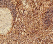
ARG66773 anti-CD44 antibody [SQab20206] IHC-P image
Immunohistochemistry: Formalin/PFA-fixed and paraffin-embedded Human tonsil tissue. Antigen Retrieval: Heat mediation was performed in Tris/EDTA buffer (pH 9.0). The tissue section was stained with ARG66773 anti-CD44 antibody [SQab20206] at 18°C - 25°C for 30 minutes.


