ARG54697
anti-CD44 antibody
anti-CD44 antibody for Flow cytometry,ICC/IF,IHC-Formalin-fixed paraffin-embedded sections,Western blot and Human
Cancer antibody; Developmental Biology antibody; Immune System antibody; Chondrogenesis Study antibody

Overview
| Product Description | Mouse Monoclonal antibody recognizes CD44 |
|---|---|
| Tested Reactivity | Hu |
| Tested Application | FACS, ICC/IF, IHC-P, WB |
| Host | Mouse |
| Clonality | Monoclonal |
| Clone | Hermes-3 |
| Isotype | IgG2a, Kappa |
| Target Name | CD44 |
| Immunogen | CD44 recombinant protein. |
| Conjugation | Un-conjugated |
| Alternate Names | MDU2; MDU3; GP90 lymphocyte homing/adhesion receptor; Hermes antigen; Extracellular matrix receptor III; PGP-I; Epican; CDW44; Phagocytic glycoprotein 1; Pgp1; HUTCH-I; MC56; Hyaluronate receptor; CD antigen CD44; Heparan sulfate proteoglycan; CD44 antigen; LHR; IN; HCELL; Phagocytic glycoprotein I; PGP-1; CSPG8; MIC4; ECMR-III; CDw44 |
Application Instructions
| Application Suggestion |
|
||||||||||
|---|---|---|---|---|---|---|---|---|---|---|---|
| Application Note | * The dilutions indicate recommended starting dilutions and the optimal dilutions or concentrations should be determined by the scientist. | ||||||||||
| Positive Control | HeLa |
Properties
| Purification | Protein G purified |
|---|---|
| Buffer | PBS and 0.09% (W/V) Sodium azide |
| Preservative | 0.09% (W/V) Sodium azide |
| Storage Instruction | For continuous use, store undiluted antibody at 2-8°C for up to a week. For long-term storage, aliquot and store at -20°C or below. Storage in frost free freezers is not recommended. Avoid repeated freeze/thaw cycles. Suggest spin the vial prior to opening. The antibody solution should be gently mixed before use. |
| Note | For laboratory research only, not for drug, diagnostic or other use. |
Bioinformation
| Database Links | |
|---|---|
| Gene Symbol | CD44 |
| Gene Full Name | CD44 molecule (Indian blood group) |
| Background | The protein encoded by this gene is a cell-surface glycoprotein involved in cell-cell interactions, cell adhesion and migration. It is a receptor for hyaluronic acid (HA) and can also interact with other ligands, such as osteopontin, collagens, and matrix metalloproteinases (MMPs). This protein participates in a wide variety of cellular functions including lymphocyte activation, recirculation and homing, hematopoiesis, and tumor metastasis. Transcripts for this gene undergo complex alternative splicing that results in many functionally distinct isoforms, however, the full length nature of some of these variants has not been determined. Alternative splicing is the basis for the structural and functional diversity of this protein, and may be related to tumor metastasis. [provided by RefSeq, Jul 2008] |
| Function | Receptor for hyaluronic acid (HA). Mediates cell-cell and cell-matrix interactions through its affinity for HA, and possibly also through its affinity for other ligands such as osteopontin, collagens, and matrix metalloproteinases (MMPs). Adhesion with HA plays an important role in cell migration, tumor growth and progression. In cancer cells, may play an important role in invadopodia formation. Also involved in lymphocyte activation, recirculation and homing, and in hematopoiesis. Altered expression or dysfunction causes numerous pathogenic phenotypes. Great protein heterogeneity due to numerous alternative splicing and post-translational modification events. [From Uniprot] |
| Cellular Localization | Cell membrane; Single-pass type I membrane protein. Note=Colocalizes with actin in membrane protrusions at wounding edges. |
| Research Area | Cancer antibody; Developmental Biology antibody; Immune System antibody; Chondrogenesis Study antibody |
| Calculated MW | 82 kDa |
| PTM | Proteolytically cleaved in the extracellular matrix by specific proteinases (possibly MMPs) in several cell lines and tumors. N- and O-glycosylated. O-glycosylation contains more-or-less-sulfated chondroitin sulfate glycans, whose number may affect the accessibility of specific proteinases to their cleavage site(s). It is uncertain if O-glycosylation occurs on Thr-637 or Thr-638. Phosphorylated; activation of PKC results in the dephosphorylation of Ser-706 (constitutive phosphorylation site), and the phosphorylation of Ser-672. |
Images (4) Click the Picture to Zoom In
-
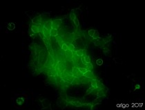
ARG54697 anti-CD44 antibody ICC/IF image
Immunofluorescence: 100% Methanol fixed (RT, 10 min) HeLa cells stained with ARG54697 anti-CD44 antibody (green) at 1:100 dilution.
Secondary antibody: ARG55393 Goat anti-Mouse IgG (H+L) antibody (FITC)
-
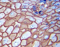
ARG54697 anti-CD44 antibody IHC-P image
Immunohistochemistry: Paraffin-embedded Human esophagus carcinoma stained with ARG54697 anti-CD44 antibody.
-
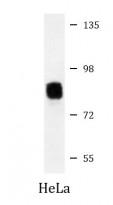
ARG54697 anti-CD44 antibody WB image
Western blot: 35 µg of HeLa cell lysate stained with ARG54697 anti-CD44 antibody.
-
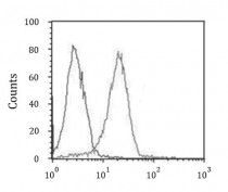
ARG54697 anti-CD44 antibody FACS image
Flow Cytometry: HeLa cells stained with ARG54697 anti-CD44 antibody (right histogram) or without primary antibody control (left histogram).


Customer's Feedback
 Excellent

Review for anti-CD44 antibody
Application:IF/ICC
Sample:HeLa
Fixation Buffer:100% Methanol
Fixation Time:10 min
Fixation Temperature:RT ºC
Permeabilization Buffer:0.1% Triton X-100
Primary Antibody Dilution Factor:1:100
Primary Antibody Incubation Time:overnight
Primary Antibody Incubation Temperature:4 ºC
Conjugation of Secondary Antibody:FITC

Excellent

Review for anti-CD44 antibody
Application:WB
Sample:HUVEC cell lysate
Sample Loading Amount:30 µg
Primary Antibody Dilution Factor:1:500
Primary Antibody Incubation Time:overnight
Primary Antibody Incubation Temperature:4 ºC
 Average

Review for anti-CD44 antibody
Application:WB
Sample:U87 cell lysate
Sample Loading Amount:30 µg
Primary Antibody Dilution Factor:1:500
Primary Antibody Incubation Time:overnight
Primary Antibody Incubation Temperature:4 ºC


