ARG23064
anti-CD43 antibody [W3/13]
anti-CD43 antibody [W3/13] for Flow cytometry,IHC-Frozen sections,IHC-Formalin-fixed paraffin-embedded sections and Rat
Overview
| Product Description | Mouse Monoclonal antibody [W3/13] recognizes CD43 Mouse anti Rat CD43 antibody, clone W3/13 recognizes the rat CD43 cell surface antigen, also known as leukosialin, sialophorin or W3/13 antigen. CD43 is a 371 amino acid ~95 kDa heavily glycosylated single pass type 1 transmebrane glycoprotein (Killeen et al. 1987) expressed by all leucocytes with the exception of B lymphocytes. CD43, in mice acts as a T-cell counter-receptor for CD169 (Siglec-1) suggesting a role in cell-cell interactions (van den Berg et al. 2001)Mouse anti Rat CD43 antibody, clone W3/13 is routinely tested in flow cytometry on rat splenocytes. |
|---|---|
| Tested Reactivity | Rat |
| Tested Application | FACS, IHC-Fr, IHC-P |
| Host | Mouse |
| Clonality | Monoclonal |
| Clone | W3/13 |
| Isotype | IgG1 |
| Target Name | CD43 |
| Antigen Species | Rat |
| Immunogen | Rat thymocyte membrane glycoproteins. |
| Conjugation | Un-conjugated |
| Alternate Names | LSN; CD43; GALGP; GPL115 |
Application Instructions
| Application Suggestion |
|
||||||||
|---|---|---|---|---|---|---|---|---|---|
| Application Note | IHC-P: This product requires antigen retrieval using heat treatment prior to staining of paraffin sections. FACS: Use 10 µl of the suggested working dilution to label 10^6 cells in 100 µl. * The dilutions indicate recommended starting dilutions and the optimal dilutions or concentrations should be determined by the scientist. |
Properties
| Form | Liquid |
|---|---|
| Purification | Purification with Protein G. |
| Buffer | PBS and 0.09% Sodium azide |
| Preservative | 0.09% Sodium azide |
| Concentration | 1 mg/ml |
| Storage Instruction | For continuous use, store undiluted antibody at 2-8°C for up to a week. For long-term storage, aliquot and store at -20°C or below. Storage in frost free freezers is not recommended. Avoid repeated freeze/thaw cycles. Suggest spin the vial prior to opening. The antibody solution should be gently mixed before use. |
| Note | For laboratory research only, not for drug, diagnostic or other use. |
Bioinformation
| Gene Symbol | Spn |
|---|---|
| Gene Full Name | sialophorin |
| Background | The protein encoded by this gene is a major sialoglycoprotein found on the surface of thymocytes, T lymphocytes, monocytes, granulocytes, and some B lymphocytes. It may be part of a physiologic ligand-receptor complex involved in T-cell activation. During T-cell activation, this protein is actively removed from the T-cell-APC (antigen-presenting cell) contact site, suggesting a negative regulatory role in adaptive immune response. [provided by RefSeq, Sep 2011] |
| Function | One of the major glycoproteins of thymocytes and T lymphocytes. Plays a role in the physicochemical properties of the T-cell surface and in lectin binding. Presents carbohydrate ligands to selectins. Has an extended rodlike structure that could protrude above the glycocalyx of the cell and allow multiple glycan chains to be accessible for binding. Is a counter-receptor for SN/Siglec-1 (By similarity). During T-cell activation is actively removed from the T-cell-APC (antigen-presenting cell) contact site thus suggesting a negative regulatory role in adaptive immune response (By similarity). [UniProt] |
| Calculated MW | 40 kDa |
Images (6) Click the Picture to Zoom In
-
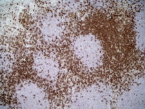
ARG23064 anti-CD43 antibody [W3/13] IHC-Fr image
Immunohistochemistry: Rat lymph node cryosection stained with ARG23064 anti-CD43 antibody [W3/13] followed by peroxidase conjugated Goat anti Mouse IgG1 antibody for detection. (Low power).
-

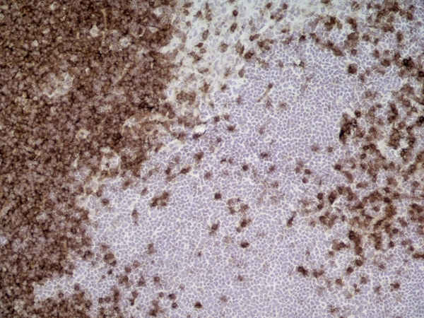
ARG23064 anti-CD43 antibody [W3/13] IHC-Fr image
Immunohistochemistry: Rat lymph node cryosection stained with ARG23064 anti-CD43 antibody [W3/13] followed by peroxidase conjugated Goat anti Mouse IgG1 antibody for detection. (Medium power).
-
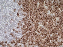
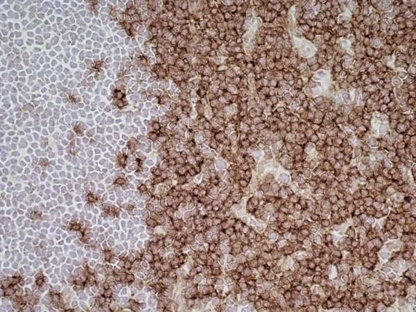
ARG23064 anti-CD43 antibody [W3/13] IHC-Fr image
Immunohistochemistry: Rat lymph node cryosection stained with ARG23064 anti-CD43 antibody [W3/13] followed by peroxidase conjugated Goat anti Mouse IgG1 antibody for detection. (High power).
-
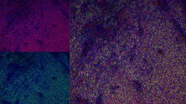
ARG23064 anti-CD43 antibody [W3/13] IHC-Fr image
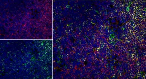
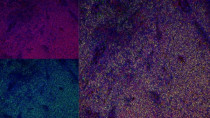
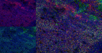
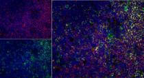
Immunohistochemistry: Rat lymph node cryosection stained with ARG23064 anti-CD43 antibody [W3/13] in red and Mouse anti Rat CD4 in green. Merged image is on the right. (Low power).
-
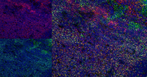
ARG23064 anti-CD43 antibody [W3/13] IHC-Fr image
Immunohistochemistry: Rat lymph node cryosection stained with ARG23064 anti-CD43 antibody [W3/13] in red and Mouse anti Rat CD4 in green. Merged image is on the right. (Medium power).
-
ARG23064 anti-CD43 antibody [W3/13] IHC-Fr image
Immunohistochemistry: Rat lymph node cryosection stained with ARG23064 anti-CD43 antibody [W3/13] in red and Mouse anti Rat CD4 in green. Merged image is on the right. (High power).