ARG62847
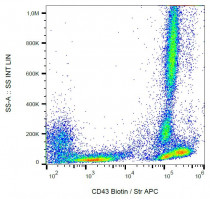
anti-CD43 antibody [MEM-59] (Biotin)
anti-CD43 antibody [MEM-59] (Biotin) for Flow cytometry and Human
Developmental Biology antibody; Immune System antibody
Overview
| Product Description | Biotin-conjugated Mouse Monoclonal antibody [MEM-59] recognizes CD43 |
|---|---|
| Tested Reactivity | Hu |
| Tested Application | FACS |
| Specificity | The clone MEM-59 recognizes neuraminidase-sensitive epitope on CD43 (Leukosialin), a 95-135 kDa type I transmembrane glycoprotein (mucin-type) which is involved in lymphocyte activation. CD43 is expressed by platelets and at high levels on the surface of all leukocytes; it is negative on resting B lymphocytes and erythrocytes. HLDA IV; WS Code NL 604 HLDA V; WS Code AS S290 |
| Host | Mouse |
| Clonality | Monoclonal |
| Clone | MEM-59 |
| Isotype | IgG1 |
| Target Name | CD43 |
| Antigen Species | Human |
| Immunogen | Human T lymphocytes. |
| Conjugation | Biotin |
| Alternate Names | CD antigen CD43; B-cell differentiation antigen LP-3; Sialophorin; Cd43; Galgp; A630014B01Rik; Ly-48; Lymphocyte antigen 48; Ly48; Leukocyte sialoglycoprotein; Leukosialin |
Application Instructions
| Application Suggestion |
|
||||
|---|---|---|---|---|---|
| Application Note | * The dilutions indicate recommended starting dilutions and the optimal dilutions or concentrations should be determined by the scientist. |
Properties
| Form | Liquid |
|---|---|
| Purification Note | The purified antibody is conjugated with Biotin-LC-NHS under optimum conditions. The reagent is free of unconjugated biotin. |
| Buffer | TBS (pH 8.0) and 15 mM Sodium azide |
| Preservative | 15 mM Sodium azide |
| Concentration | 1 mg/ml |
| Storage Instruction | Aliquot and store in the dark at 2-8°C. Keep protected from prolonged exposure to light. Avoid repeated freeze/thaw cycles. Suggest spin the vial prior to opening. The antibody solution should be gently mixed before use. |
| Note | For laboratory research only, not for drug, diagnostic or other use. |
Bioinformation
| Database Links | |
|---|---|
| Gene Symbol | SPN |
| Gene Full Name | sialophorin |
| Background | CD43 (leukosialin, sialophorin) is a transmembrane mucin-like protein with high negative charge, expressed on the surface of most hematopoietic cells. CD43 contributes to a repulsive barrier that interferes with cellular adhesion, however, in certain cases also promotes leukocyte aggregation. By interaction with actin-binding proteins ezrin and moesin CD43 plays a regulatory role in remodeling T-cell morphology and regulates cell-cell interactions during lymphocyte traffic. CD43 signaling both enhances LFA-1 adhesiveness and counteracts LFA-1 induction via other receptors. Expression of CD43 causes induction of functionally active tumour suppressor p53 protein, but in case of p53 and ARF defficiency CD43 promotes tumour proliferation and viability. It appears to be an important modulator of leukocyte functions. |
| Function | One of the major glycoproteins of thymocytes and T lymphocytes. Plays a role in the physicochemical properties of the T-cell surface and in lectin binding. Presents carbohydrate ligands to selectins. Has an extended rodlike structure that could protrude above the glycocalyx of the cell and allow multiple glycan chains to be accessible for binding. Is a counter-receptor for SN/Siglec-1 (By similarity). During T-cell activation is actively removed from the T-cell-APC (antigen-presenting cell) contact site thus suggesting a negative regulatory role in adaptive immune response (By similarity). [UniProt] |
| Research Area | Developmental Biology antibody; Immune System antibody |
| Calculated MW | 40 kDa |
Images (1) Click the Picture to Zoom In
Clone References


