ARG42224
anti-CD43 antibody [4I3]
anti-CD43 antibody [4I3] for Flow cytometry,IHC-Formalin-fixed paraffin-embedded sections,Western blot and Human,Mouse,Rat
Overview
| Product Description | Mouse Monoclonal antibody [4I3] recognizes CD43 |
|---|---|
| Tested Reactivity | Hu, Ms, Rat |
| Tested Application | FACS, IHC-P, WB |
| Host | Mouse |
| Clonality | Monoclonal |
| Clone | 4I3 |
| Isotype | IgG |
| Target Name | CD43 |
| Antigen Species | Human |
| Immunogen | Recombinant protein corresponding to A272-P400 of Human CD43. |
| Conjugation | Un-conjugated |
| Alternate Names | LSN; CD43; GALGP; GPL115 |
Application Instructions
| Application Suggestion |
|
||||||||
|---|---|---|---|---|---|---|---|---|---|
| Application Note | IHC-P: Antigen Retrieval: Heat mediation was performed in Citrate buffer (pH 6.0, epitope retrieval solution) for 20 min. * The dilutions indicate recommended starting dilutions and the optimal dilutions or concentrations should be determined by the scientist. |
||||||||
| Observed Size | ~ 115 kDa |
Properties
| Form | Liquid |
|---|---|
| Purification | Affinity purification with immunogen. |
| Buffer | 0.2% Na2HPO4, 0.9% NaCl, 0.05% Sodium azide and 4% Trehalose. |
| Preservative | 0.05% Sodium azide |
| Stabilizer | 4% Trehalose |
| Concentration | 0.5 mg/ml |
| Storage Instruction | For continuous use, store undiluted antibody at 2-8°C for up to a week. For long-term storage, aliquot and store at -20°C or below. Storage in frost free freezers is not recommended. Avoid repeated freeze/thaw cycles. Suggest spin the vial prior to opening. The antibody solution should be gently mixed before use. |
| Note | For laboratory research only, not for drug, diagnostic or other use. |
Bioinformation
| Database Links | |
|---|---|
| Gene Symbol | SPN |
| Gene Full Name | sialophorin |
| Background | This gene encodes a highly sialylated glycoprotein that functions in antigen-specific activation of T cells, and is found on the surface of thymocytes, T lymphocytes, monocytes, granulocytes, and some B lymphocytes. It contains a mucin-like extracellular domain, a transmembrane region and a carboxy-terminal intracellular region. The extracellular domain has a high proportion of serine and threonine residues, allowing extensive O-glycosylation, and has one potential N-glycosylation site, while the carboxy-terminal region has potential phosphorylation sites that may mediate transduction of activation signals. Different glycoforms of this protein have been described. In stimulated immune cells, proteolytic cleavage of the extracellular domain occurs in some cell types, releasing a soluble extracellular fragment. Defects in expression of this gene are associated with Wiskott-Aldrich syndrome. [provided by RefSeq, Sep 2017] |
| Function | Predominant cell surface sialoprotein of leukocytes which regulates multiple T-cell functions, including T-cell activation, proliferation, differentiation, trafficking and migration. Positively regulates T-cell trafficking to lymph-nodes via its association with ERM proteins (EZR, RDX and MSN) (By similarity). Negatively regulates Th2 cell differentiation and predisposes the differentiation of T-cells towards a Th1 lineage commitment. Promotes the expression of IFN-gamma by T-cells during T-cell receptor (TCR) activation of naive cells and induces the expression of IFN-gamma by CD4+ T-cells and to a lesser extent by CD8+ T-cells (PubMed:18036228). Plays a role in preparing T-cells for cytokine sensing and differentiation into effector cells by inducing the expression of cytokine receptors IFNGR and IL4R, promoting IFNGR and IL4R signaling and by mediating the clustering of IFNGR with TCR (PubMed:24328034). Acts as a major E-selectin ligand responsible for Th17 cell rolling on activated vasculature and recruitment during inflammation. Mediates Th17 cells, but not Th1 cells, adhesion to E-selectin. Acts as a T-cell counter-receptor for SIGLEC1. [UniProt] |
| Cellular Localization | Membrane; Single-pass type I membrane protein. Cell projection, microvillus. Cell projection, uropodium. Note=Localizes to the uropodium and microvilli via its interaction with ERM proteins (EZR, RDX and MSN). CD43 cytoplasmic tail: Nucleus. Nucleus, PML body. [UniProt] |
| Calculated MW | 40 kDa |
Images (7) Click the Picture to Zoom In
-

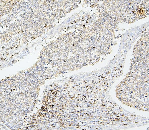
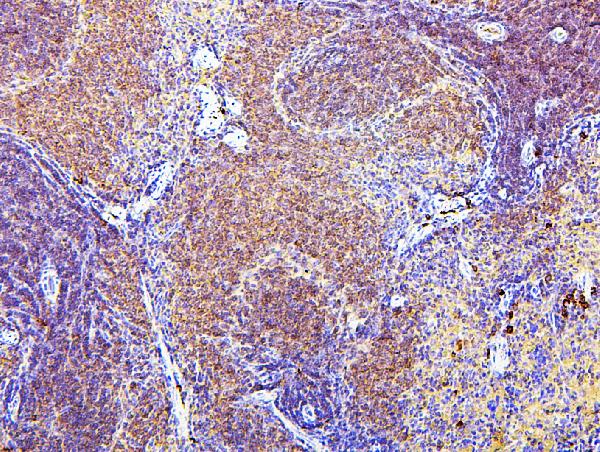
ARG42224 anti-CD43 antibody [4I3] IHC-P image
Immunohistochemistry: Paraffin-embedded Human lung cancer tissue. Antigen Retrieval: Heat mediation was performed in Citrate buffer (pH 6.0, epitope retrieval solution) for 20 min. The tissue section was blocked with 10% goat serum. The tissue section was then stained with ARG42224 anti-CD43 antibody [4I3] at 1 µg/ml dilution, overnight at 4°C.
-
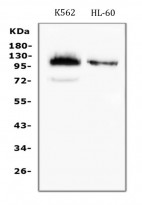
ARG42224 anti-CD43 antibody [4I3] WB image
Western blot: 50 µg of samples under reducing conditions. K562 and HL-60 whole cell lysates stained with ARG42224 anti-CD43 antibody [4I3] at 0.5 µg/ml dilution, overnight at 4°C.
-
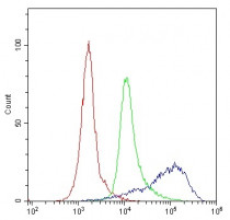
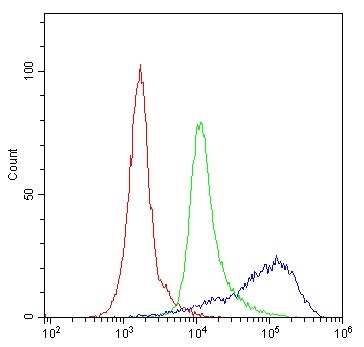
ARG42224 anti-CD43 antibody [4I3] FACS image
Flow Cytometry: Human PBMC cells were blocked with 10% normal goat serum and then stained with ARG42224 anti-CD43 antibody [4I3] (blue line) at 1 µg/10^6 cells for 30 min at 20°C, followed by incubation with DyLight®488 labelled secondary antibody. Isotype control antibody (green line) was Mouse IgG (1 µg/10^6 cells) used under the same conditions. Unlabelled sample (red line) was also used as a control.
-
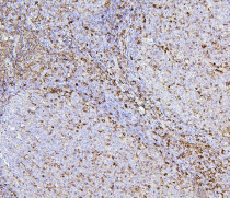
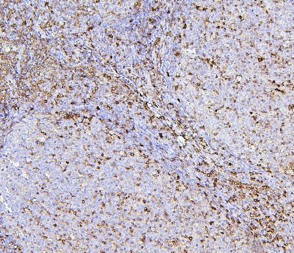
ARG42224 anti-CD43 antibody [4I3] IHC-P image
Immunohistochemistry: Paraffin-embedded Human tonsil tissue. Antigen Retrieval: Heat mediation was performed in Citrate buffer (pH 6.0, epitope retrieval solution) for 20 min. The tissue section was blocked with 10% goat serum. The tissue section was then stained with ARG42224 anti-CD43 antibody [4I3] at 1 µg/ml dilution, overnight at 4°C.
-
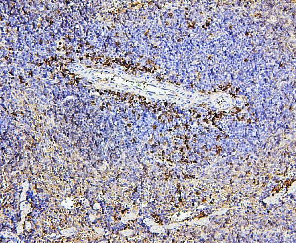
ARG42224 anti-CD43 antibody [4I3] IHC-P image
Immunohistochemistry: Paraffin-embedded Mouse spleen tissue. Antigen Retrieval: Heat mediation was performed in Citrate buffer (pH 6.0, epitope retrieval solution) for 20 min. The tissue section was blocked with 10% goat serum. The tissue section was then stained with ARG42224 anti-CD43 antibody [4I3] at 1 µg/ml dilution, overnight at 4°C.
-

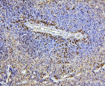
ARG42224 anti-CD43 antibody [4I3] IHC-P image
Immunohistochemistry: Paraffin-embedded Rat spleen tissue. Antigen Retrieval: Heat mediation was performed in Citrate buffer (pH 6.0, epitope retrieval solution) for 20 min. The tissue section was blocked with 10% goat serum. The tissue section was then stained with ARG42224 anti-CD43 antibody [4I3] at 1 µg/ml dilution, overnight at 4°C.