ARG23028
anti-CD36 antibody [MF3] (PE)
anti-CD36 antibody [MF3] (PE) for Flow cytometry and Mouse
Overview
| Product Description | PE-conjugated Rat Monoclonal antibody [MF3] recognizes CD36 Rat anti Mouse CD36 antibody, clone MF3 recognizes mouse CD36, also known as platelet glycoprotein 4, glycoprotein IIIb or PAS IV. CD36 is an ~85 kDa multipass transmembrane glycoprotein primarily expressed on platelets, monocytes/macrophages, smooth muscle and endothelial cells. The CD36 molecule is type B scavenger receptor, which binds to multiple ligands including thrombospondin, anionic phospholipids, oxidized low density lipoproteins and long chain fatty acids. CD36 has diverse functions and is reported to play a role in innate immunity, platelet adhesion/aggregation and long chain fatty acid transport. The CD36 molecule also directly mediates cytoadhesion of erythrocytes infected with Plasmodium falciparum, and may be involved in the development of atherosclerotic lesions and the formation of foam cells.Rat anti Mouse CD36 antibody, clone MF3 has been shown to inhibit IL-4 induced thioglycollate-elicited peritoneal macrophage fusion and significantly block IL-4/GM-CSF-induced bone-marrorw derived macrophage fusion. |
|---|---|
| Tested Reactivity | Ms |
| Tested Application | FACS |
| Host | Rat |
| Clonality | Monoclonal |
| Clone | MF3 |
| Isotype | IgG2a |
| Target Name | CD36 |
| Antigen Species | Mouse |
| Immunogen | IL-4 treated murine thioglycollate-elicited peritoneal macrophages |
| Conjugation | PE |
| Alternate Names | GPIV; CHDS7; Platelet glycoprotein 4; CD antigen CD36; PAS-4; PASIV; Glycoprotein IIIb; PAS IV; GPIIIB; FAT; SCARB3; GP3B; Leukocyte differentiation antigen CD36; Platelet collagen receptor; BDPLT10; Thrombospondin receptor; GP4; Fatty acid translocase; Platelet glycoprotein IV |
Application Instructions
| Application Suggestion |
|
||||
|---|---|---|---|---|---|
| Application Note | FACS: Use 10 µl of the suggested working dilution to label 10^6 cells in 100 µl. * The dilutions indicate recommended starting dilutions and the optimal dilutions or concentrations should be determined by the scientist. |
Properties
| Form | Liquid |
|---|---|
| Purification | Purification with Protein G. |
| Buffer | PBS, 0.09% Sodium azide, 1% BSA and 5% Sucrose |
| Preservative | 0.09% Sodium azide |
| Stabilizer | 1% BSA and 5% Sucrose |
| Storage Instruction | Aliquot and store in the dark at 2-8°C. Keep protected from prolonged exposure to light. Avoid repeated freeze/thaw cycles. Suggest spin the vial prior to opening. The antibody solution should be gently mixed before use. |
| Note | For laboratory research only, not for drug, diagnostic or other use. |
Bioinformation
| Database Links | |
|---|---|
| Gene Symbol | Cd36 |
| Gene Full Name | CD36 antigen |
| Background | The protein encoded by this gene is the fourth major glycoprotein of the platelet surface and serves as a receptor for thrombospondin in platelets and various cell lines. Since thrombospondins are widely distributed proteins involved in a variety of adhesive processes, this protein may have important functions as a cell adhesion molecule. It binds to collagen, thrombospondin, anionic phospholipids and oxidized LDL. It directly mediates cytoadherence of Plasmodium falciparum parasitized erythrocytes and it binds long chain fatty acids and may function in the transport and/or as a regulator of fatty acid transport. Mutations in this gene cause platelet glycoprotein deficiency. Multiple alternatively spliced transcript variants have been found for this gene. [provided by RefSeq, Feb 2014] |
| Function | Binds to collagen, thrombospondin, anionic phospholipids and oxidized low-density lipoprotein (oxLDL). May function as a cell adhesion molecule. Directly mediates cytoadherence of Plasmodium falciparum parasitized erythrocytes. Binds long chain fatty acids and may function in the transport and/or as a regulator of fatty acid transport. Receptor for thombospondins, THBS1 AND THBS2, mediating their antiangiogenic effects. As a coreceptor for TLR4-TLR6 heterodimer, promotes inflammation in monocytes/macrophages. Upon ligand binding, such as oxLDL or amyloid-beta 42, rapidly induces the formation of a heterodimer of TLR4 and TLR6, which is internalized and triggers inflammatory response, leading to NF-kappa-B-dependent production of CXCL1, CXCL2 and CCL9 cytokines, via MYD88 signaling pathway, and CCL5 cytokine, via TICAM1 signaling pathway, as well as IL1B secretion. [UniProt] |
| Calculated MW | 53 kDa |
| PTM | N-glycosylated and O-glycosylated with a ratio of 2:1. Ubiquitinated at Lys-469 and Lys-472. Ubiquitination is induced by fatty acids such as oleic acid and leads to degradation by the proteasome (PubMed:21610069, PubMed:18353783). Ubiquitination and degradation are inhibited by insulin which blocks the effect of fatty acids (PubMed:18353783). |
Images (1) Click the Picture to Zoom In
-
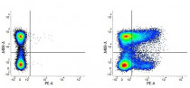
ARG23028 anti-CD36 antibody [MF3] (PE) FACS image
Flow Cytometry: Figure A. A488-conjugated Rat anti Mouse CD45R and PE-conjugated Rat IgG2a isotype control. Figure B. A488 conjugated Rat anti Mouse CD45R and ARG23028 anti-CD36 antibody [MF3] (PE). All experiments performed on Mouse splenocytes.


