ARG66342
anti-CD31 antibody [SQab18116]
anti-CD31 antibody [SQab18116] for Flow cytometry,IHC-Formalin-fixed paraffin-embedded sections,Immunoprecipitation,Western blot and Human
Cancer antibody; Cell Biology and Cellular Response antibody; Controls and Markers antibody; Developmental Biology antibody; Signaling Transduction antibody; Endothelial Cell Marker antibody; Microvascular Density Study antibody
Overview
| Product Description | Recombinant Rabbit Monoclonal antibody [SQab18116] recognizes CD31 |
|---|---|
| Tested Reactivity | Hu |
| Tested Application | FACS, IHC-P, IP, WB |
| Host | Rabbit |
| Clonality | Monoclonal |
| Clone | SQab18116 |
| Isotype | IgG |
| Target Name | CD31 |
| Antigen Species | Human |
| Immunogen | Synthetic peptide corresponding to residues on the C-terminus of Human CD31. |
| Conjugation | Un-conjugated |
| Alternate Names | EndoCAM; CD31/EndoCAM; PECAM-1; CD31; PECA1; CD antigen CD31; GPIIA'; endoCAM; Platelet endothelial cell adhesion molecule |
Application Instructions
| Application Suggestion |
|
||||||||||
|---|---|---|---|---|---|---|---|---|---|---|---|
| Application Note | IHC-P: Antigen Retrieval: Heat mediated was performed using Tris/EDTA buffer (pH 9.0). * The dilutions indicate recommended starting dilutions and the optimal dilutions or concentrations should be determined by the scientist. |
||||||||||
| Observed Size | ~ 140 kDa |
Properties
| Form | Liquid |
|---|---|
| Purification | Purification with Protein A. |
| Buffer | PBS, 0.01% Sodium azide, 40% Glycerol and 0.05% BSA. |
| Preservative | 0.01% Sodium azide |
| Stabilizer | 40% Glycerol and 0.05% BSA |
| Storage Instruction | For continuous use, store undiluted antibody at 2-8°C for up to a week. For long-term storage, aliquot and store at -20°C. Storage in frost free freezers is not recommended. Avoid repeated freeze/thaw cycles. Suggest spin the vial prior to opening. The antibody solution should be gently mixed before use. |
| Note | For laboratory research only, not for drug, diagnostic or other use. |
Bioinformation
| Database Links |
Swiss-port # P16284 Human Platelet endothelial cell adhesion molecule |
|---|---|
| Gene Symbol | PECAM1 |
| Gene Full Name | platelet/endothelial cell adhesion molecule 1 |
| Background | CD31 protein is found on the surface of platelets, monocytes, neutrophils, and some types of T-cells, and makes up a large portion of endothelial cell intercellular junctions. The encoded protein is a member of the immunoglobulin superfamily and is likely involved in leukocyte migration, angiogenesis, and integrin activation. [provided by RefSeq, May 2010] |
| Function | CD31 is a cell adhesion molecule which is required for leukocyte transendothelial migration (TEM) under most inflammatory conditions (PubMed:19342684, PubMed:17580308). Tyr-690 plays a critical role in TEM and is required for efficient trafficking of PECAM1 to and from the lateral border recycling compartment (LBRC) and is also essential for the LBRC membrane to be targeted around migrating leukocytes (PubMed:19342684). Trans-homophilic interaction may play a role in endothelial cell-cell adhesion via cell junctions (PubMed:27958302). Heterophilic interaction with CD177 plays a role in transendothelial migration of neutrophils (PubMed:17580308). Homophilic ligation of PECAM1 prevents macrophage-mediated phagocytosis of neighboring viable leukocytes by transmitting a detachment signal (PubMed:12110892). Promotes macrophage-mediated phagocytosis of apoptotic leukocytes by tethering them to the phagocytic cells; PECAM1-mediated detachment signal appears to be disabled in apoptotic leukocytes (PubMed:12110892). Modulates bradykinin receptor BDKRB2 activation (PubMed:18672896). Regulates bradykinin- and hyperosmotic shock-induced ERK1/2 activation in endothelial cells (PubMed:18672896). Induces susceptibility to atherosclerosis. Isoform Delta15: Does not protect against apoptosis. [UniProt] |
| Highlight | Related products: CD31 antibodies; CD31 ELISA Kits; CD31 Duos / Panels; Anti-Rabbit IgG secondary antibodies; Related news: Cancer Pathology Markers (SQ clones) |
| Research Area | Cancer antibody; Cell Biology and Cellular Response antibody; Controls and Markers antibody; Developmental Biology antibody; Signaling Transduction antibody; Endothelial Cell Marker antibody; Microvascular Density Study antibody |
| Calculated MW | 83 kDa |
| PTM | Phosphorylated on Ser and Tyr residues after cellular activation. Phosphorylated on tyrosine residues by FER and FES in response to FCER1 activation (By similarity). In endothelial cells Fyn mediates mechanical-force (stretch or pull) induced tyrosine phosphorylation. Palmitoylation by ZDHHC21 is necessary for cell surface expression in endothelial cells and enrichment in membrane rafts. [UniProt] |
Images (5) Click the Picture to Zoom In
-
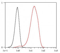
ARG66342 anti-CD31 antibody [SQab18116] FACS image
Flow Cytometry: Jurkat cells fixed with 4% paraformaldehyde for 10 min. The cells were then stained with ARG66342 anti-CD31 antibody [SQab18116] (red) at 1:1000 dilution in 1x PBS/1% BSA for 30 min at room temperature, followed by Alexa Fluor® 488 labelled secondary antibody. Unlabelled sample (black) was used as a control.
-
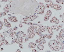
ARG66342 anti-CD31 antibody [SQab18116] IHC-P image
Immunohistochemistry: Formalin-fixed and paraffin-embedded Human placenta stained with ARG66342 anti-CD31 antibody [SQab18116] at 1:3200 dilution. Antigen Retrieval: Heat mediated was performed using Tris/EDTA buffer (pH 9.0).
-
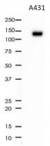
ARG66342 anti-CD31 antibody [SQab18116] WB image
Western blot: 10 µg of A431 cell lysate stained with ARG66342 anti-CD31 antibody [SQab18116] at 1:1000 dilution.
-
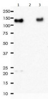
ARG66342 anti-CD31 antibody [SQab18116] IP image
Immunoprecipitation: 0.4 mg of THP-1 whole cell lysate was immunoprecipitated (1:50 dilution) and stained with ARG66342 anti-CD31 antibody [SQab18116].
Lane 1: Immunoprecipitation in THP-1 whole cell lysate
Lane 2: PBS instead of Primary Ab in THP-1 whole cell lysate
Lane 3: THP-1 whole cell lysate, 10 µg (input) -
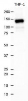
ARG66342 anti-CD31 antibody [SQab18116] WB image
Western blot: 10 µg of THP-1 cell lysate stained with ARG66342 anti-CD31 antibody [SQab18116] at 1:1000 dilution.


Specific References
PD-L1 and VEGF dual blockade enhances anti-tumor effect on brain metastasis in hematogenous metastasis model
IHC-P / Mouse


BRAF V600E mutant colorectal cancer cells mediate local immunosuppressive microenvironment through exosomal long noncoding RNAs
IHC-P / Human


