ARG56119
anti-CD31 antibody [JC/70A]
anti-CD31 antibody [JC/70A] for Flow cytometry,ICC/IF,IHC-Formalin-fixed paraffin-embedded sections,Western blot and Human,Mouse
Cancer antibody; Cell Biology and Cellular Response antibody; Controls and Markers antibody; Developmental Biology antibody; Signaling Transduction antibody; Endothelial Cell Marker antibody; Microvascular Density Study antibody

Overview
| Product Description | Mouse Monoclonal antibody [JC/70A] recognizes CD31 |
|---|---|
| Tested Reactivity | Hu, Ms |
| Predict Reactivity | Rb |
| Species Does Not React With | Rat, Pig |
| Tested Application | FACS, ICC/IF, IHC-P, WB |
| Host | Mouse |
| Clonality | Monoclonal |
| Clone | JC/70A |
| Isotype | IgG1, kappa |
| Target Name | CD31 |
| Antigen Species | Human |
| Immunogen | A membrane preparation of a spleen from a patient with hairy cell leukemia. |
| Conjugation | Un-conjugated |
| Alternate Names | EndoCAM; CD31/EndoCAM; PECAM-1; CD31; PECA1; CD antigen CD31; GPIIA'; endoCAM; Platelet endothelial cell adhesion molecule |
Application Instructions
| Predict Reactivity Note | Duo to the publication PMID: 35242369, this antibody might react to rabbit CD31. | ||||||||||
|---|---|---|---|---|---|---|---|---|---|---|---|
| Application Suggestion |
|
||||||||||
| Application Note | IHC-P: Antigen Retrieval: Boil tissue section in 10 mM Tris with 1 mM EDTA (pH 9.0) for 10-20 min, followed by cooling at RT for 20 min. * The dilutions indicate recommended starting dilutions and the optimal dilutions or concentrations should be determined by the scientist. |
||||||||||
| Positive Control | HUVEC | ||||||||||
| Observed Size | 110 - 120 kDa |
Properties
| Form | Liquid |
|---|---|
| Purification | Purification with Protein G. |
| Buffer | PBS (pH 7.4), 0.05% Sodium azide and 0.1 mg/ml BSA |
| Preservative | 0.05% Sodium azide |
| Stabilizer | 0.1 mg/ml BSA |
| Concentration | 0.2 mg/ml |
| Storage Instruction | For continuous use, store undiluted antibody at 2-8°C for up to a week. For long-term storage, aliquot and store at -20°C or below. Storage in frost free freezers is not recommended. Avoid repeated freeze/thaw cycles. Suggest spin the vial prior to opening. The antibody solution should be gently mixed before use. |
| Note | For laboratory research only, not for drug, diagnostic or other use. |
Bioinformation
| Database Links |
Swiss-port # P16284 Human Platelet endothelial cell adhesion molecule Swiss-port # Q08481 Mouse Platelet endothelial cell adhesion molecule |
|---|---|
| Gene Symbol | PECAM1 |
| Gene Full Name | platelet/endothelial cell adhesion molecule 1 |
| Background | CD31 protein is found on the surface of platelets, monocytes, neutrophils, and some types of T-cells, and makes up a large portion of endothelial cell intercellular junctions. The encoded protein is a member of the immunoglobulin superfamily and is likely involved in leukocyte migration, angiogenesis, and integrin activation. [provided by RefSeq, May 2010] |
| Function | CD31 is a cell adhesion molecule which is required for leukocyte transendothelial migration (TEM) under most inflammatory conditions (PubMed:19342684, PubMed:17580308). Tyr-690 plays a critical role in TEM and is required for efficient trafficking of PECAM1 to and from the lateral border recycling compartment (LBRC) and is also essential for the LBRC membrane to be targeted around migrating leukocytes (PubMed:19342684). Trans-homophilic interaction may play a role in endothelial cell-cell adhesion via cell junctions (PubMed:27958302). Heterophilic interaction with CD177 plays a role in transendothelial migration of neutrophils (PubMed:17580308). Homophilic ligation of PECAM1 prevents macrophage-mediated phagocytosis of neighboring viable leukocytes by transmitting a detachment signal (PubMed:12110892). Promotes macrophage-mediated phagocytosis of apoptotic leukocytes by tethering them to the phagocytic cells; PECAM1-mediated detachment signal appears to be disabled in apoptotic leukocytes (PubMed:12110892). Modulates bradykinin receptor BDKRB2 activation (PubMed:18672896). Regulates bradykinin- and hyperosmotic shock-induced ERK1/2 activation in endothelial cells (PubMed:18672896). Induces susceptibility to atherosclerosis. Isoform Delta15: Does not protect against apoptosis. [UniProt] |
| Cellular Localization | Cell surface and cytoplasm of endothelial cells |
| Highlight | Related products: CD31 antibodies; CD31 Duos / Panels; Anti-Mouse IgG secondary antibodies; Related news: Besides tumor suppression, what’s p53 busy for during embryogenesis? |
| Research Area | Cancer antibody; Cell Biology and Cellular Response antibody; Controls and Markers antibody; Developmental Biology antibody; Signaling Transduction antibody; Endothelial Cell Marker antibody; Microvascular Density Study antibody |
| Calculated MW | 83 kDa |
| PTM | Phosphorylated on Ser and Tyr residues after cellular activation. Phosphorylated on tyrosine residues by FER and FES in response to FCER1 activation (By similarity). In endothelial cells Fyn mediates mechanical-force (stretch or pull) induced tyrosine phosphorylation. Palmitoylation by ZDHHC21 is necessary for cell surface expression in endothelial cells and enrichment in membrane rafts. |
Images (4) Click the Picture to Zoom In
-
_210_205.jpg)
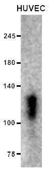
ARG56119 anti-CD31 antibody [JC/70A] WB image
Western blot: 10 µg of HUVEC cell lysate stained with ARG56119 anti-CD31 antibody [JC/70A] at 1:200 dilution.
-
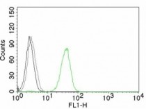
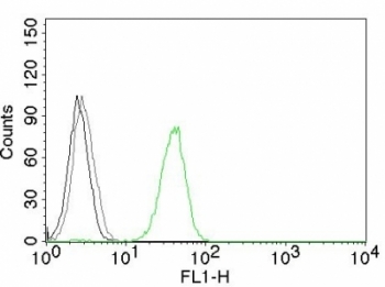
ARG56119 anti-CD31 antibody [JC/70A] FACS image
Flow Cytometry: Jurkat cells stained with AF488-conjugated ARG56119 anti-CD31 antibody [JC/70A] (green); Cells alone (black); Isotype control (grey).
-
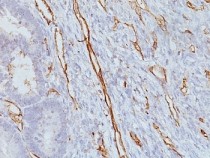
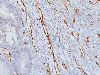
ARG56119 anti-CD31 antibody [JC/70A] IHC-P image
Immunohistochemistry: Formalin-fixed, paraffin-embedded Human colon carcinoma stained with ARG56119 anti-CD31 antibody [JC/70A].
-
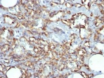
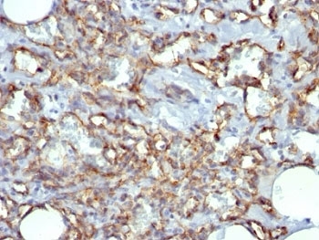
ARG56119 anti-CD31 antibody [JC/70A] IHC-P image
Immunohistochemistry: Formalin-fixed, paraffin-embedded Human angiosarcoma stained with ARG56119 anti-CD31 antibody [JC/70A].
.jpg)
Customer's Feedback
Excellent
Review for anti-CD31 antibody [JC/70A]
Application:WB
Sample:HUVEC
Sample Loading Amount:10 µg
Primary Antibody Dilution Factor:1:200
Primary Antibody Incubation Time:overnight
Primary Antibody Incubation Temperature:4 ºC

Specific References
CT perfusion imaging can detect residual lung tumor early after radiofrequency ablation: a preliminary animal study on both tumoral and peri-tumoral region assessment
IHC-P / Rabbit


