ARG59393
anti-CD2AP antibody
anti-CD2AP antibody for Western blot and Human,Mouse,Rat
Overview
| Product Description | Rabbit Polyclonal antibody recognizes CD2AP |
|---|---|
| Tested Reactivity | Hu, Ms, Rat |
| Tested Application | WB |
| Host | Rabbit |
| Clonality | Polyclonal |
| Isotype | IgG |
| Target Name | CD2AP |
| Antigen Species | Human |
| Immunogen | Synthetic peptide of Human CD2AP |
| Conjugation | Un-conjugated |
| Alternate Names | Adapter protein CMS; CD2-associated protein; CMS; Cas ligand with multiple SH3 domains |
Application Instructions
| Application Suggestion |
|
||||
|---|---|---|---|---|---|
| Application Note | * The dilutions indicate recommended starting dilutions and the optimal dilutions or concentrations should be determined by the scientist. | ||||
| Positive Control | A431 | ||||
| Observed Size | 71 kDa |
Properties
| Form | Liquid |
|---|---|
| Purification | Affinity purified. |
| Buffer | PBS (pH 7.3), 0.02% Sodium azide and 50% Glycerol. |
| Preservative | 0.02% Sodium azide |
| Stabilizer | 50% Glycerol |
| Storage Instruction | For continuous use, store undiluted antibody at 2-8°C for up to a week. For long-term storage, aliquot and store at -20°C. Storage in frost free freezers is not recommended. Avoid repeated freeze/thaw cycles. Suggest spin the vial prior to opening. The antibody solution should be gently mixed before use. |
| Note | For laboratory research only, not for drug, diagnostic or other use. |
Bioinformation
| Database Links | |
|---|---|
| Gene Symbol | CD2AP |
| Gene Full Name | CD2-associated protein |
| Background | This gene encodes a scaffolding molecule that regulates the actin cytoskeleton. The protein directly interacts with filamentous actin and a variety of cell membrane proteins through multiple actin binding sites, SH3 domains, and a proline-rich region containing binding sites for SH3 domains. The cytoplasmic protein localizes to membrane ruffles, lipid rafts, and the leading edges of cells. It is implicated in dynamic actin remodeling and membrane trafficking that occurs during receptor endocytosis and cytokinesis. Haploinsufficiency of this gene is implicated in susceptibility to glomerular disease. [provided by RefSeq, Jul 2008] |
| Function | Seems to act as an adapter protein between membrane proteins and the actin cytoskeleton. In collaboration with CBLC, modulates the rate of RET turnover and may act as regulatory checkpoint that limits the potency of GDNF on neuronal survival. Controls CBLC function, converting it from an inhibitor to a promoter of RET degradation. May play a role in receptor clustering and cytoskeletal polarity in the junction between T-cell and antigen-presenting cell. May anchor the podocyte slit diaphragm to the actin cytoskeleton in renal glomerolus. Also required for cytokinesis. [UniProt] |
| Cellular Localization | Cytoplasm, cytoskeleton. Cell projection, ruffle. Cell junction. Note=Colocalizes with F-actin and BCAR1/p130Cas in membrane ruffles (PubMed:10339567). Located at podocyte slit diaphragm between podocyte foot processes (By similarity). During late anaphase and telophase, concentrates in the vicinity of the midzone microtubules and in the midbody in late telophase (PubMed:15800069). [UniProt] |
| Calculated MW | 71 kDa |
| PTM | Phosphorylated on tyrosine residues; probably by c-Abl, Fyn and c-Src. [UniProt] |
Images (1) Click the Picture to Zoom In
-

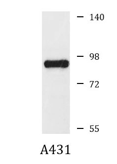
ARG59393 anti-CD2AP antibody WB image
Western blot: 25 µg of A431 cell lysate stained with ARG59393 anti-CD2AP antibody at 1:1000 dilution.