ARG59346
anti-CD2AP antibody [5F8]
anti-CD2AP antibody [5F8] for Flow cytometry,ICC/IF,IHC-Formalin-fixed paraffin-embedded sections,Western blot and Human,Mouse,Rat
Overview
| Product Description | Mouse Monoclonal antibody [5F8] recognizes CD2AP |
|---|---|
| Tested Reactivity | Hu, Ms, Rat |
| Tested Application | FACS, ICC/IF, IHC-P, WB |
| Host | Mouse |
| Clonality | Monoclonal |
| Clone | 5F8 |
| Isotype | IgG1 |
| Target Name | CD2AP |
| Antigen Species | Human |
| Immunogen | Recombinant protein corresponding to K253-K337 of Human CD2AP. |
| Conjugation | Un-conjugated |
| Alternate Names | Adapter protein CMS; CD2-associated protein; CMS; Cas ligand with multiple SH3 domains |
Application Instructions
| Application Suggestion |
|
||||||||||
|---|---|---|---|---|---|---|---|---|---|---|---|
| Application Note | IHC-P: Antigen Retrieval: Heat mediation was performed in Citrate buffer (pH 6.0) for 20 min. * The dilutions indicate recommended starting dilutions and the optimal dilutions or concentrations should be determined by the scientist. |
Properties
| Form | Liquid |
|---|---|
| Purification | Affinity purification with immunogen. |
| Buffer | 0.9% NaCl, 0.2% Na2HPO4, 0.05% Sodium azide and 4% Trehalose. |
| Preservative | 0.05% Sodium azide |
| Stabilizer | 4% Trehalose |
| Concentration | 0.5 mg/ml |
| Storage Instruction | For continuous use, store undiluted antibody at 2-8°C for up to a week. For long-term storage, aliquot and store at -20°C or below. Storage in frost free freezers is not recommended. Avoid repeated freeze/thaw cycles. Suggest spin the vial prior to opening. The antibody solution should be gently mixed before use. |
| Note | For laboratory research only, not for drug, diagnostic or other use. |
Bioinformation
| Database Links | |
|---|---|
| Gene Symbol | CD2AP |
| Gene Full Name | CD2-associated protein |
| Background | This gene encodes a scaffolding molecule that regulates the actin cytoskeleton. The protein directly interacts with filamentous actin and a variety of cell membrane proteins through multiple actin binding sites, SH3 domains, and a proline-rich region containing binding sites for SH3 domains. The cytoplasmic protein localizes to membrane ruffles, lipid rafts, and the leading edges of cells. It is implicated in dynamic actin remodeling and membrane trafficking that occurs during receptor endocytosis and cytokinesis. Haploinsufficiency of this gene is implicated in susceptibility to glomerular disease. [provided by RefSeq, Jul 2008] |
| Function | Seems to act as an adapter protein between membrane proteins and the actin cytoskeleton. In collaboration with CBLC, modulates the rate of RET turnover and may act as regulatory checkpoint that limits the potency of GDNF on neuronal survival. Controls CBLC function, converting it from an inhibitor to a promoter of RET degradation. May play a role in receptor clustering and cytoskeletal polarity in the junction between T-cell and antigen-presenting cell. May anchor the podocyte slit diaphragm to the actin cytoskeleton in renal glomerolus. Also required for cytokinesis. [UniProt] |
| Cellular Localization | Cytoplasm, cytoskeleton. Cell projection, ruffle. Cell junction. Note=Colocalizes with F-actin and BCAR1/p130Cas in membrane ruffles (PubMed:10339567). Located at podocyte slit diaphragm between podocyte foot processes (By similarity). During late anaphase and telophase, concentrates in the vicinity of the midzone microtubules and in the midbody in late telophase (PubMed:15800069). [UniProt] |
| Calculated MW | 71 kDa |
| PTM | Phosphorylated on tyrosine residues; probably by c-Abl, Fyn and c-Src. [UniProt] |
Images (12) Click the Picture to Zoom In
-

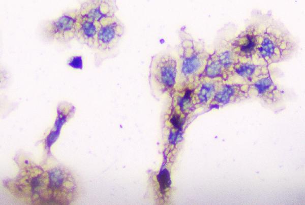
ARG59346 anti-CD2AP antibody [5F8] ICC image
Immunocytochemistry: A431 cells were blocked with 10% goat serum and then stained with ARG59346 anti-CD2AP antibody [5F8] at 1 µg/ml dilution, overnight at 4°C.
-
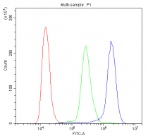
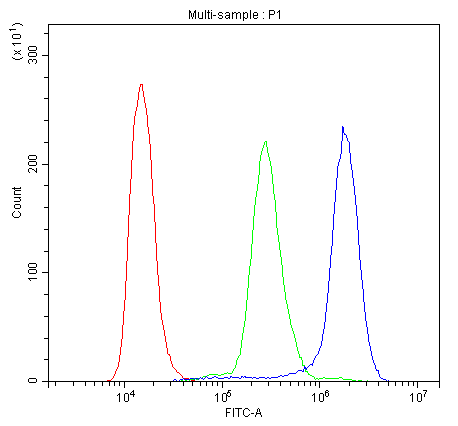
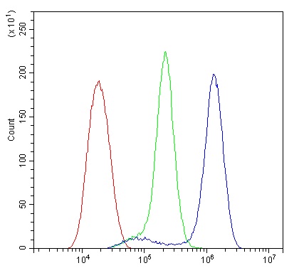
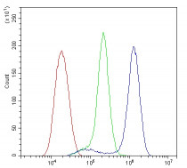
ARG59346 anti-CD2AP antibody [5F8] FACS image
Flow Cytometry: K562 cells were blocked with 10% normal goat serum and then stained with ARG59346 anti-CD2AP antibody [5F8] (blue) at 1 µg/10^6 cells for 30 min at 20°C, followed by DyLight®488 labelled secondary antibody. Isotype control antibody (green) was Mouse IgG (1 µg/10^6 cells) used under the same conditions. Unlabelled sample (red) was also used as a control.
-
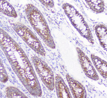
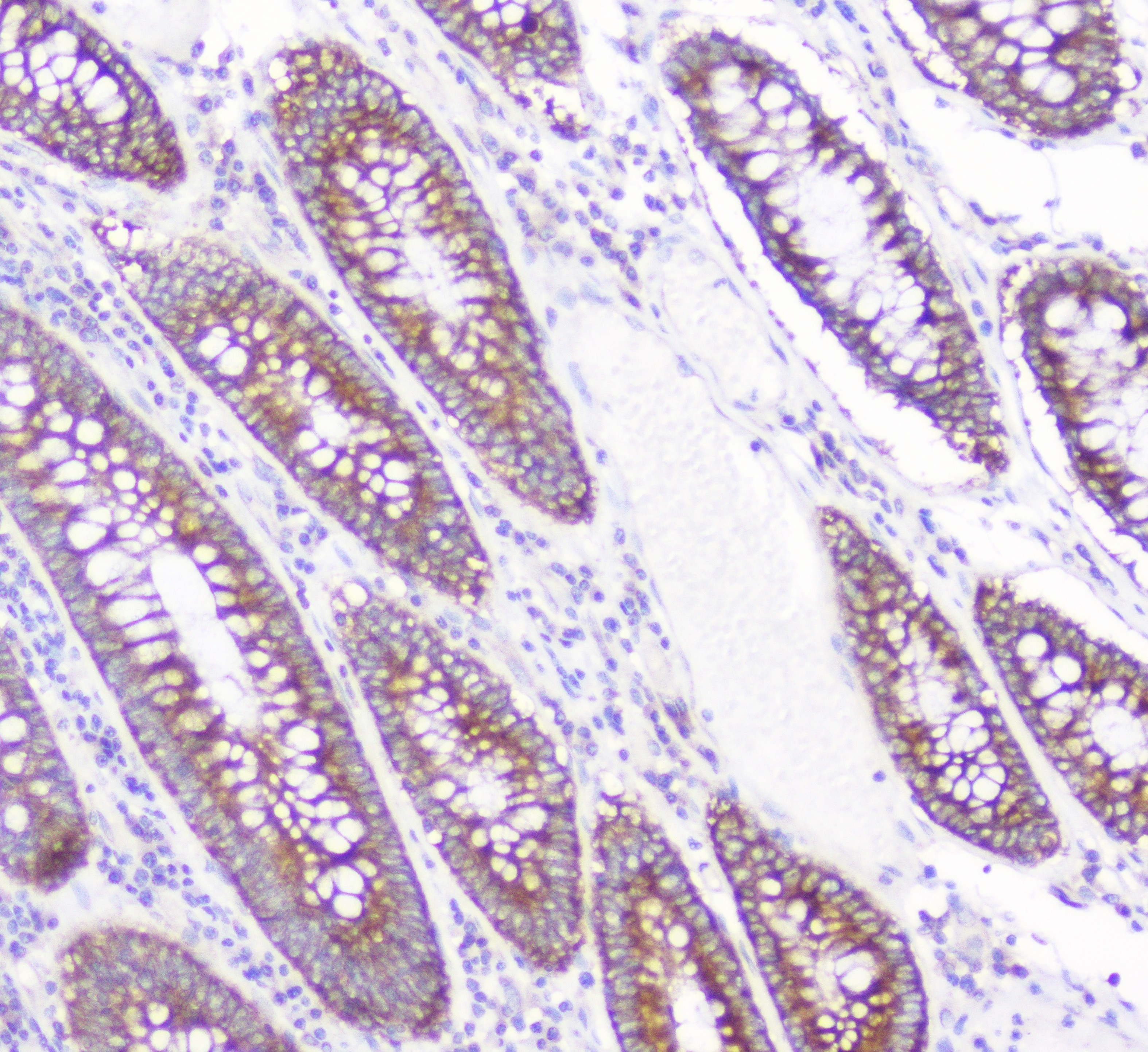
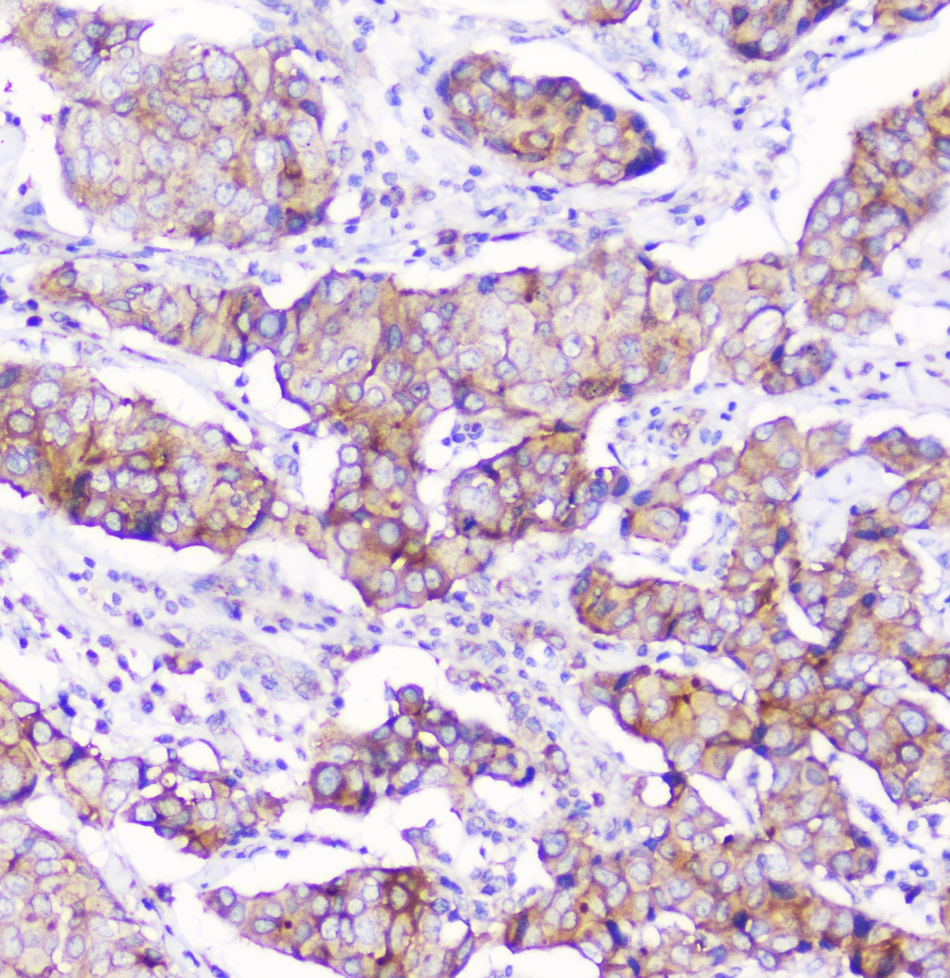
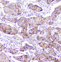
ARG59346 anti-CD2AP antibody [5F8] IHC-P image
Immunohistochemistry: Paraffin-embedded Human colon cancer. Antigen Retrieval: Heat mediated was performed in Citrate buffer (pH 6.0, epitope retrieval solution) for 20 min. The tissue section was blocked with 10% goat serum. The tissue section was then stained with ARG59346 anti-CD2AP antibody [5F8] at 1 µg/ml dilution, overnight at 4°C.
-
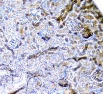
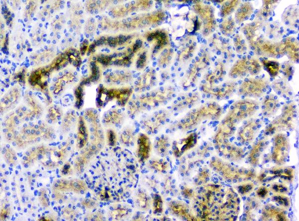
ARG59346 anti-CD2AP antibody [5F8] IHC-P image
Immunohistochemistry: Paraffin-embedded Mouse kidney tissue. Antigen Retrieval: Heat mediation was performed in Citrate buffer (pH 6.0) for 20 min. The tissue section was blocked with 10% goat serum. The tissue section was then stained with ARG59346 anti-CD2AP antibody [5F8] at 1 µg/ml dilution, overnight at 4°C.
-

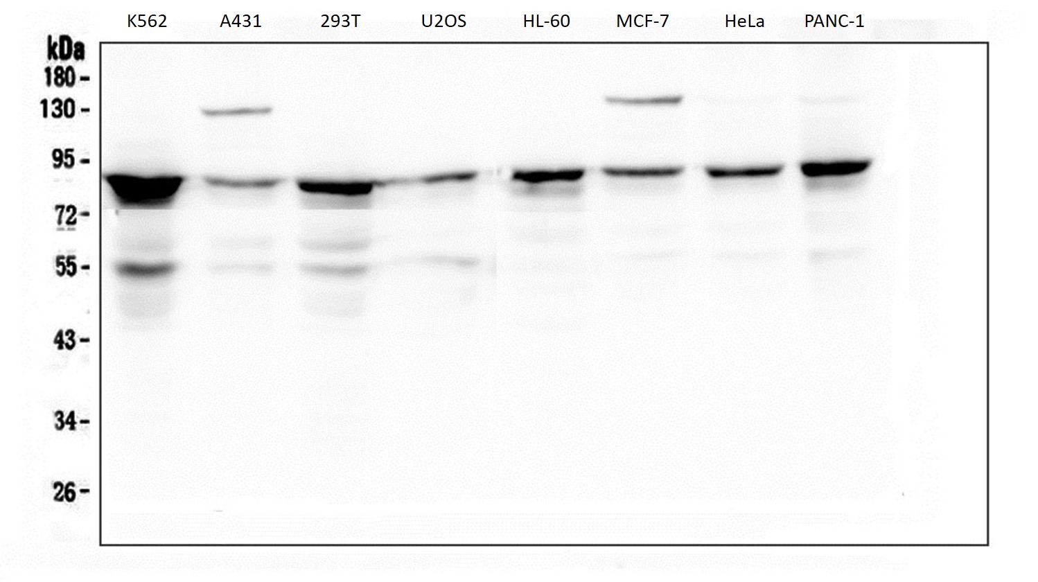
ARG59346 anti-CD2AP antibody [5F8] WB image
Western blot: 50 µg of samples under reducing conditions. K562, A431, 293T, U2OS, HL-60, MCF-7, HeLa and PANC-1 cell lysates stained with ARG59346 anti-CD2AP antibody [5F8] at 0.5 µg/ml, overnight at 4°C.
-
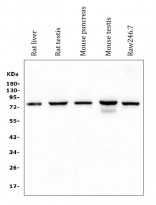
ARG59346 anti-CD2AP antibody [5F8] WB image
Western blot: 50 µg of sample under reducing conditions. Rat liver, Rat testis, Mouse pancreas, Mouse testis and Raw246.7 whole cell lysates stained with ARG59346 anti-CD2AP antibody [5F8] at 0.5 µg/ml dilution, overnight at 4°C.
-
ARG59346 anti-CD2AP antibody [5F8] FACS image
Flow Cytometry: U2OS cells were blocked with 10% normal goat serum and then stained with ARG59346 anti-CD2AP antibody [5F8] (blue) at 1 µg/10^6 cells for 30 min at 20°C, followed by incubation with DyLight®488 labelled secondary antibody. Isotype control antibody (green) was rabbit IgG (1 µg/10^6 cells) used under the same conditions. Unlabelled sample (red) was also used as a control.
-
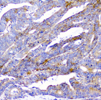
ARG59346 anti-CD2AP antibody [5F8] IHC-P image
Immunohistochemistry: Paraffin-embedded Human colon cancer. Antigen Retrieval: Heat mediated was performed in Citrate buffer (pH 6.0, epitope retrieval solution) for 20 min. The tissue section was blocked with 10% goat serum. The tissue section was then stained with ARG59346 anti-CD2AP antibody [5F8] at 1 µg/ml dilution, overnight at 4°C.
-
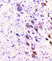
ARG59346 anti-CD2AP antibody [5F8] IHC-P image
Immunohistochemistry: Paraffin-embedded Rat brain tissue. Antigen Retrieval: Heat mediation was performed in Citrate buffer (pH 6.0) for 20 min. The tissue section was blocked with 10% goat serum. The tissue section was then stained with ARG59346 anti-CD2AP antibody [5F8] at 1 µg/ml dilution, overnight at 4°C.
-
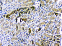
ARG59346 anti-CD2AP antibody [5F8] IHC-P image
Immunohistochemistry: Paraffin-embedded Rat kidney tissue. Antigen Retrieval: Heat mediation was performed in Citrate buffer (pH 6.0) for 20 min. The tissue section was blocked with 10% goat serum. The tissue section was then stained with ARG59346 anti-CD2AP antibody [5F8] at 1 µg/ml dilution, overnight at 4°C.
-
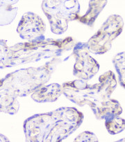
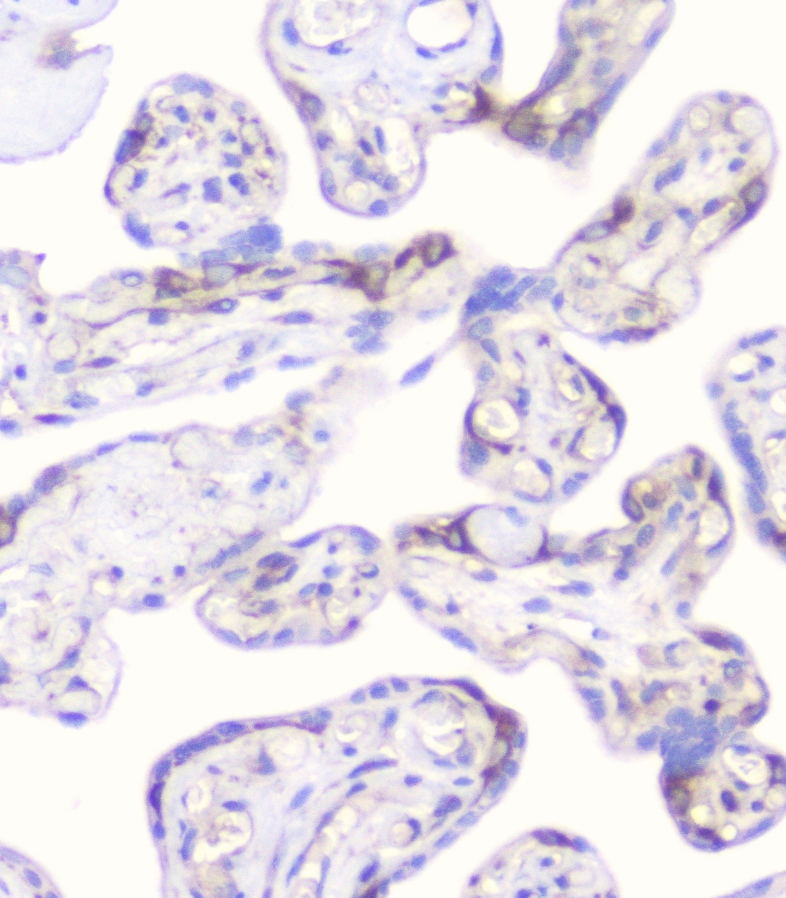
ARG59346 anti-CD2AP antibody [5F8] IHC-P image
Immunohistochemistry: Paraffin-embedded Human placenta tissue. Antigen Retrieval: Heat mediated was performed in Citrate buffer (pH 6.0, epitope retrieval solution) for 20 min. The tissue section was blocked with 10% goat serum. The tissue section was then stained with ARG59346 anti-CD2AP antibody [5F8] at 1 µg/ml dilution, overnight at 4°C.
-
ARG59346 anti-CD2AP antibody [5F8] IHC-P image
Immunohistochemistry: Paraffin-embedded Human mammary cancer. Antigen Retrieval: Heat mediated was performed in Citrate buffer (pH 6.0, epitope retrieval solution) for 20 min. The tissue section was blocked with 10% goat serum. The tissue section was then stained with ARG59346 anti-CD2AP antibody [5F8] at 1 µg/ml dilution, overnight at 4°C.