ARG54263
anti-CD235a antibody [JC159] (PE)
anti-CD235a antibody [JC159] (PE) for Flow cytometry and Human,Rat
Cell Biology and Cellular Response antibody
Overview
| Product Description | PE-conjugated Mouse Monoclonal antibody [JC159] recognizes CD235a |
|---|---|
| Tested Reactivity | Hu, Rat |
| Tested Application | FACS |
| Specificity | The mouse monoclonal antibody JC159 recognizes an epitope between amino acids 27 and 40 of the extracellular portion of CD235a (glycophorin A), a sialoglycoprotein expressed on early erythroblasts, late erythroblasts, erythroblasts, mature erythrocytes and the cells of erythroid cell lines K562 and HEL. The antibody does not react with glycophorin B. |
| Host | Mouse |
| Clonality | Monoclonal |
| Clone | JC159 |
| Isotype | IgG1 |
| Target Name | CD235a |
| Immunogen | Membrane preparation from splenic hairy cell leukemia |
| Conjugation | PE |
| Alternate Names | MN; GPErik; MNS; GPA; GPSAT; PAS-2; MN sialoglycoprotein; CD235a; HGpMiV; CD antigen CD235a; HGpMiXI; Sialoglycoprotein alpha; HGpSta(C); Glycophorin-A |
Application Instructions
| Application Suggestion |
|
||||
|---|---|---|---|---|---|
| Application Note | * The dilutions indicate recommended starting dilutions and the optimal dilutions or concentrations should be determined by the scientist. |
Properties
| Form | Liquid |
|---|---|
| Purification Note | The purified antibody is conjugated with R-Phycoerythrin (PE) under optimum conditions. The conjugate is purified by size-exclusion chromatography and adjusted for direct use. No reconstitution is necessary. |
| Buffer | PBS, 15 mM Sodium azide and 0.2% (w/v) high-grade protease free BSA |
| Preservative | 15 mM Sodium azide |
| Stabilizer | 0.2% (w/v) high-grade protease free BSA |
| Storage Instruction | Aliquot and store in the dark at 2-8°C. Keep protected from prolonged exposure to light. Avoid repeated freeze/thaw cycles. Suggest spin the vial prior to opening. The antibody solution should be gently mixed before use. |
| Note | For laboratory research only, not for drug, diagnostic or other use. |
Bioinformation
| Database Links | |
|---|---|
| Gene Symbol | GYPA |
| Gene Full Name | glycophorin A (MNS blood group) |
| Background | CD235a (Glycophorin A, GPA) is a transmembrane sialoglycoprotein expressed on erythrocytes and their precursors. Similarly to glycophorin B (GPB), these molecules provide the cells with a large mucin-like surface, which minimalizes aggregation between erythrocytes in the circulation. GPA is the carrier of blood group M and N specificities, while GPB accounts for S, s and U specificities. CD235a is a receptor of Hsa, an Streptococcus adhesin. |
| Function | Glycophorin A is the major intrinsic membrane protein of the erythrocyte. The N-terminal glycosylated segment, which lies outside the erythrocyte membrane, has MN blood group receptors. Appears to be important for the function of SLC4A1 and is required for high activity of SLC4A1. May be involved in translocation of SLC4A1 to the plasma membrane. Is a receptor for influenza virus. Is a receptor for Plasmodium falciparum erythrocyte-binding antigen 175 (EBA-175); binding of EBA-175 is dependent on sialic acid residues of the O-linked glycans. Appears to be a receptor for Hepatitis A virus (HAV). [UniProt] |
| Research Area | Cell Biology and Cellular Response antibody |
| Calculated MW | 16 kDa |
| PTM | The major O-linked glycan are NeuAc-alpha-(2-3)-Gal-beta-(1-3)-[NeuAc-alpha-(2-6)]-GalNAcOH (about 78 %) and NeuAc-alpha-(2-3)-Gal-beta-(1-3)-GalNAcOH (17 %). Minor O-glycans (5 %) include NeuAc-alpha-(2-3)-Gal-beta-(1-3)-[NeuAc-alpha-(2-6)]-GalNAcOH NeuAc-alpha-(2-8)-NeuAc-alpha-(2-3)-Gal-beta-(1-3)-GalNAcOH. About 1% of all O-linked glycans carry blood group A, B and H determinants. They derive from a type-2 precursor core structure, Gal-beta-(1,3)-GlcNAc-beta-1-R, and the antigens are synthesized by addition of fucose (H antigen-specific) and then N-acetylgalactosamine (A antigen-specific) or galactose (B antigen-specific). Specifically O-linked-glycans are NeuAc-alpha-(2-3)-Gal-beta-(1-3)-GalNAcOH-(6-1)-GlcNAc-beta-(4-1)-[Fuc-alpha-(1-2)]-Gal-beta-(3-1)-GalNAc-alpha (about 1%, B antigen-specific) and NeuAc-alpha-(2-3)-Gal-beta-(1-3)-GalNAcOH-(6-1)-GlcNAc-beta-(4-1)-[Fuc-alpha-(1-2)]-Gal-beta (1 %, O antigen-, A antigen- and B antigen-specific). |
Images (2) Click the Picture to Zoom In
-

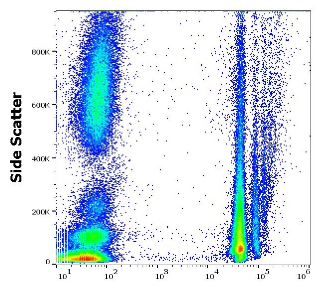
ARG54263 anti-CD235a antibody [JC159] (PE) FACS image
Flow Cytometry: Human peripheral whole blood stained with ARG54263 anti-CD235a antibody [JC159] (PE) (10 µl reagent / 100 µl of peripheral whole blood).
-
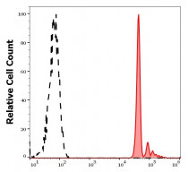
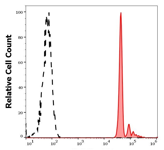
ARG54263 anti-CD235a antibody [JC159] (PE) FACS image
Flow Cytometry: Separation of human erythrocytes (red-filled) from neutrophil granulocytes (black-dashed). Human peripheral whole blood stained with ARG54263 anti-CD235a antibody [JC159] (PE) (10 µl reagent / 100 µl of peripheral whole blood).
Clone References
Genome-wide analysis reveals that Smad3 and JMJD3 HDM co-activate the neural developmental program.


