ARG41462
anti-CD235a antibody
anti-CD235a antibody for Flow cytometry,IHC-Formalin-fixed paraffin-embedded sections,Western blot and Human
Overview
| Product Description | Rabbit Polyclonal antibody recognizes CD235a |
|---|---|
| Tested Reactivity | Hu |
| Tested Application | FACS, IHC-P, WB |
| Host | Rabbit |
| Clonality | Polyclonal |
| Isotype | IgG |
| Target Name | CD235a |
| Antigen Species | Human |
| Immunogen | Synthetic peptide of Human CD235a. |
| Conjugation | Un-conjugated |
| Alternate Names | MN; GPErik; MNS; GPA; GPSAT; PAS-2; MN sialoglycoprotein; CD235a; HGpMiV; CD antigen CD235a; HGpMiXI; Sialoglycoprotein alpha; HGpSta(C); Glycophorin-A |
Application Instructions
| Application Suggestion |
|
||||||||
|---|---|---|---|---|---|---|---|---|---|
| Application Note | * The dilutions indicate recommended starting dilutions and the optimal dilutions or concentrations should be determined by the scientist. | ||||||||
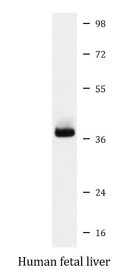
| Positive Control | Human fetal liver | ||||||||
| Observed Size | ~ 37 kDa |
Properties
| Form | Liquid |
|---|---|
| Purification | Affinity purified. |
| Buffer | PBS (pH 7.4), 150 mM NaCl, 0.02% Sodium azide and 50% Glycerol. |
| Preservative | 0.02% Sodium azide |
| Stabilizer | 50% Glycerol |
| Storage Instruction | For continuous use, store undiluted antibody at 2-8°C for up to a week. For long-term storage, aliquot and store at -20°C. Storage in frost free freezers is not recommended. Avoid repeated freeze/thaw cycles. Suggest spin the vial prior to opening. The antibody solution should be gently mixed before use. |
| Note | For laboratory research only, not for drug, diagnostic or other use. |
Bioinformation
| Database Links | |
|---|---|
| Gene Symbol | GYPA |
| Gene Full Name | glycophorin A (MNS blood group) |
| Background | Glycophorins A (GYPA) and B (GYPB) are major sialoglycoproteins of the human erythrocyte membrane which bear the antigenic determinants for the MN and Ss blood groups. In addition to the M or N and S or s antigens that commonly occur in all populations, about 40 related variant phenotypes have been identified. These variants include all the variants of the Miltenberger complex and several isoforms of Sta, as well as Dantu, Sat, He, Mg, and deletion variants Ena, S-s-U- and Mk. Most of the variants are the result of gene recombinations between GYPA and GYPB. [provided by RefSeq, Jul 2008] |
| Function | Glycophorin A is the major intrinsic membrane protein of the erythrocyte. The N-terminal glycosylated segment, which lies outside the erythrocyte membrane, has MN blood group receptors. Appears to be important for the function of SLC4A1 and is required for high activity of SLC4A1. May be involved in translocation of SLC4A1 to the plasma membrane. Is a receptor for influenza virus. Is a receptor for Plasmodium falciparum erythrocyte-binding antigen 175 (EBA-175); binding of EBA-175 is dependent on sialic acid residues of the O-linked glycans. Appears to be a receptor for Hepatitis A virus (HAV). [UniProt] |
| Cellular Localization | Cell membrane; Single-pass type I membrane protein. Note=Appears to be colocalized with SLC4A1. [UniProt] |
| Calculated MW | 16 kDa |
| PTM | The major O-linked glycan are NeuAc-alpha-(2-3)-Gal-beta-(1-3)-[NeuAc-alpha-(2-6)]-GalNAcOH (about 78 %) and NeuAc-alpha-(2-3)-Gal-beta-(1-3)-GalNAcOH (17 %). Minor O-glycans (5 %) include NeuAc-alpha-(2-3)-Gal-beta-(1-3)-[NeuAc-alpha-(2-6)]-GalNAcOH NeuAc-alpha-(2-8)-NeuAc-alpha-(2-3)-Gal-beta-(1-3)-GalNAcOH. About 1% of all O-linked glycans carry blood group A, B and H determinants. They derive from a type-2 precursor core structure, Gal-beta-(1,3)-GlcNAc-beta-1-R, and the antigens are synthesized by addition of fucose (H antigen-specific) and then N-acetylgalactosamine (A antigen-specific) or galactose (B antigen-specific). Specifically O-linked-glycans are NeuAc-alpha-(2-3)-Gal-beta-(1-3)-GalNAcOH-(6-1)-GlcNAc-beta-(4-1)-[Fuc-alpha-(1-2)]-Gal-beta-(3-1)-GalNAc-alpha (about 1%, B antigen-specific) and NeuAc-alpha-(2-3)-Gal-beta-(1-3)-GalNAcOH-(6-1)-GlcNAc-beta-(4-1)-[Fuc-alpha-(1-2)]-Gal-beta (1 %, O antigen-, A antigen- and B antigen-specific). [UniProt] |
Images (1) Click the Picture to Zoom In
-
ARG41462 anti-CD235a antibody WB image
Western blot: Human fetal liver lysate stained with ARG41462 anti-CD235a antibody.